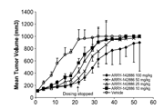
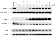
-
生物活性
HM-71224 is an EGFR tyrosine kinase inhibitor. HM-71224is an Epidermal growth factor receptor (EGFR) antagonist. Olmutinib (HM61713, HM-71224 or BI 1482694) is an oral, third-generation irreversibleEGFR mutant-selective EGFR inhibitor, for the treatment of patients withlocally advanced or metastatic EGFR T790M mutation-positive non-small cell lungcancer.
Invitro activities of olmutinib[1]
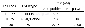
-
体外研究
-
体内研究
-
激酶实验
Inhibition test for activities of EGFR WT andL858R/T790M kinase[2]
The inhibiting activities against EGFR WTand EGFR L858R/T790M kinase were determined using z-lyte kinase assay kit. Olmutinib was prepared to 10mM DMSO solution, and a solution containing 4% DMSO wereprepared therefrom and diluted to a concentration of 1μM to 0.0001μM. Then, an approximateKd value of each kinase was calculated, and diluted using a kinase buffer (50mMHEPES (PH 7.4), 10mM MgCl2, 1mMEGTA and 0.01 % BRIJ-35) to 1 to100ng/assay concentration. The test was conducted in a 384 well polystyreneflat-bottomed plates. 5μl of the diluted solution of olmutinib was added to each well, and 10μl of a mixture of peptide substrate and kinase in a suitableconcentration and 5μl of 5~300μM ATP solution were successively added theretoand the plate was incubated in a stirrer at room temperature for 60 minutes.After 60mins, 10μl of coloring reagent was added to the resulting mixture toinitiate a fluorescence reaction of peptide substrate and a terminatingsolution was added thereto for terminating the reaction. A fluorescence valueof each well was determined with a fluorescence meter (Molecular Device) at 400nm (excitation filter) and 520nm (emission filter). The inhibiting activity ofthe test compounds against the kinases was determined as a phosphorylationpercentage (%) compared with control group, according to the kit protocol, andmeasured for IC50, the concentration of x-axis at which 50% inhibition wasobserved.
-
细胞实验
Inhibition test for growth of cancer cell expressingEGFR[2]
The inhibiting test of the inventivecompounds on the cancer cell growth was conducted in A431 (ATCC CRL-1555),HCC827 (ATCC CRL-2868) and NCI-HI975 (ATCC CRL-5908) cell lines.
A431 cell line was incubated in ahigh-glucose DMEM (Dulbecco's Modified Eagle's Medium) supplemented with 10%fetal bovine serum (FBS) and 1% penicillin/streptomycin, and HCC827 andNCI-H1975 cell lines were incubated in an RPMI medium supplemented with 10% FBS, 1 % penicillin/streptomycin and 1 % sodium pyruvate.
The cancer cell lines stored in a liquidnitrogen tank were each quickly thawed at 37oC, and centrifuged toremove the medium. The resulting cell pellet was mixed with a culture medium,incubated in a culture flask at 37oC under 5% CO2 for 2to 3 days, and the medium was removed. The remaining cells were washed withDPBS (Dulbecco's Phosphate Buffered Saline) and separated from the flask byusing Tripsin-EDTA. The separated cells were diluted with a culture medium to aconcentration of 1x10⁵ A431 cells/ml, except that in case of HCC827and NCI-HI975 cells, the dilution was carried out to 5 x10⁴cells/ml.100μ1 of the diluted cell solution was added to each well of a 96-well plate,and incubated at 37oC under 5% CO2 for 1 day. NCI-H1975cells were starved in a RPMI-1640 medium containing 0.1% FBS and 1% penicillin/streptomycinto maximize the reacting activities of the cell on the test compounds on thefollowing day.
The olmutinib wasdissolved in 99.5% dimethylsulfoxide (DMSO) to a concentration of 25mM. In casethat the test compound was not soluble in DMSO, 1 % HCl was added thereto and treatedin a 40oC water bath for 30 mins until a complete dissolution was attained.The DMSO solution containing olmutinib was diluted with a culturemedium to a final concentration of 100μM, and then diluted 10 timesserially to 10-6μM (a final concentration of DMSO was less than 1 %).
The medium was removed from each well ofthe 96-well plate. And then, 100μl of the olmutinib solution was added to each well holding the cultured cells, and the plate wasincubated at 37oC under 5% CO2 for 72 hours (except thatNCI-H1975 cells were incubated for 48 hours). After removing the medium fromthe plate, 50μl of 10% trichloroacetic acid was added to each well , and theplate was kept at 4oC for 1 hour to fix the cells to the bottom of theplate. The added 10% trichloroacetic acid solution was removed from each well,the plate was dried, 100μl of an SRB (Sulforhodamine-B) dye solution at aconcentration of 0.4% dissolved in 1% acetic acid was added thereto, and the resultingmixture was reacted for 10 mins at room temperature. After removing the dyesolution, the plate was washed with water, and well dried. When the dyesolution was not effectively removed by water, 1% acetic acid was used. 150μlof 10mM trisrna base was added to each well, and the absorbance at 540nmwavelength was determined with a microplate reader. In case of NCI-H1975, thecell viabilities were determined as the absorbance at 490 nm wavelength usingCelltiter 96 Aqueous One solution.
-
动物实验
Anticancer efficacy test in nude micexenografted with NCIH1975 cancer cells[2]
Nude mice were subcutaneous injection with1 x 108 cells/0.3 mL of NCI-HI975 cell (lung cancer cell) suspensionon the back to form of tumor.
In the test, a tumor in the sixthgeneration isolated from an individual was cut into a size of 30 mg, andtransplanted subcutaneously into right f1anks of mice using a 12-gauge trocar.The volume of tumor (V) is calculated from following equation: V= Lx S2/2after measuring a long diameter (L) and a short diameter (S) using a verniercaliper twice a week for 18 days of test. All test materials were orally administeredone time a day for total 10 days, and the tumor growth inhibition rate (lR:tumor growth inhibition rate (%) calculated based on a vehicle-treated control)and the maximum body weight loss (mBWL: maximum body weight loss calculatedbased on the body weight just before administration) were calculated usingfollowing equations: IR (%) = (1-(RTG of the treatment group of testmaterial)/(RTG of the control group)) x 100 and mBWL (%)=(l-(mean body weighton day x/ mean body weight just before administration)) x 100 wherein, RTG is arelative tumor growth, which is the mean tumor volume on a particular day basedon daily mean tumor volume, day x is a day on which the body weight loss islargest during the test.
Inhibitionon collagen- induced arthritis in mice[2]
Olmutinib was subjected to arthritis inhibitiontest in a collagen-induced arthritis (CIA) model.
Male DBA/1J mice (8 weeks old) were firstimmunized by intradermal injection of 0.7mL of a suspension liquid in which anequal volume of 2mg/mL of type II collagen is emulsified in 4mg/mL of completeFreund's adjuvant supplemented with bacteria tuberculosis. After 21 days, themice were second immunized by the injection as above, except for using asuspension liquid in which an equal volume of 2mg/mL of type II collagen isemulsified in incomplete Freund's adjuvant containing no bacteria tuberculosis.After 1 week of second immunization, mice were evaluated for clinical scoresbased on Table 10 and seven animals were grouped such that the average of experimental group isbetween 1 and 2. Test samples and vehicle of given concentrations were orally administeredin an amount of 10mL per body weight for 14 days everyday by using a Sonde. Theclinical scores of arthritis were evaluated three times a day.

-
不同实验动物依据体表面积的等效剂量转换表(数据来源于FDA指南)
|  动物 A (mg/kg) = 动物 B (mg/kg)×动物 B的Km系数/动物 A的Km系数 |
|
例如,已知某工具药用于小鼠的剂量为88 mg/kg , 则用于大鼠的剂量换算方法:将88 mg/kg 乘以小鼠的Km系数(3),再除以大鼠的Km系数(6),得到该药物用于大鼠的等效剂量44 mg/kg。
-
参考文献
[1] Kim ES. Olmutinib: First Global Approval. Drugs. 2016;76(11):1153-1157.
[more]
分子式
C26H26N6O2S |
分子量
486.59 |
CAS号
1353550-13-6 |
储存方式
﹣20 ℃冷藏长期储存。冰袋运输 |
溶剂(常温)
|
DMSO
50 mg/mL |
Water
|
Ethanol
|
体内溶解度
-
Clinical Trial Information ( data from http://clinicaltrials.gov )
注:以上所有数据均来自公开文献,并不保证对所有实验均有效,数据仅供参考。
-
相关化合物库
-
使用AMQUAR产品发表文献后请联系我们


