-
生物活性
KU-0063794 exhibits high specificity for mTOR (FRAP) inhibition. KU-0063794 at 30 nM is sufficient to rapidly ablate S6K1 activity by blocking the phosphorylation of the hydrophobic motif (Thr389) and subsequently the phosphorylation of the T-loop residue (Thr229), in HEK-293 cells. KU-0063794 at 100-300 nM completely inhibits the amino-acid-induced phosphorylation of S6K1 and S6 protein. KU-0063794 inhibits the phosphorylation of mTORC1 at Ser2448 and mTORC2 at Ser2481 in a dose-dependent and time-dependent manner. KU-0063794 induces a dose-dependent inhibition of the activity and phosphorylation of Akt at Ser473 and unexpected Thr308 as well as the phosphorylation of the Akt substrates PRAS40 at Thr246, GSK3α/GSK3β at Ser21/Ser9 and Foxo-1/3a at Thr24/Thr32, in the presence of serum or following IGF1 stimulation.
-
体外研究
-
体内研究
30% PEG400+0.5% Tween80+5% propylene glycol
-
激酶实验
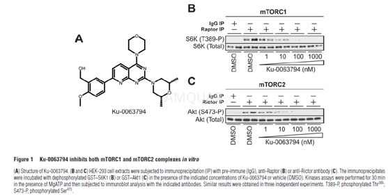
mTOR complexes kinase assays[1]
HEK-293 cells were freshly lysed in Hepeslysis buffer. Lysate (1–4 mg) was pre-cleared by incubating with 5–20μl ofProtein G–Sepharose conjugated to pre-immune IgG. The lysate extracts were thenincubated with 5–20 μl of Protein G–Sepharose conjugated to 5–20 μg of eitheranti-Rictor or anti-Raptor antibody, or pre-immune IgG. All antibodies werecovalently conjugated to Protein G–Sepharose. Immunoprecipitations were carriedout for 1 h at 4◦C on a vibrating platform. The immunoprecipitates were washedfour times with Hepes lysis buffer, followed by two washes with Hepes kinasebuffer. For Raptor immunoprecipitates used for phosphorylating S6K1, for theinitial two wash steps the buffer included 0.5 M NaCl to ensure optimal kinaseactivity. GST–Akt1 was isolated from serum-deprived HEK-293 cells incubatedwith PI-103 (1 μM for 1 h). GST–S6K1 was purified from serumdeprived HEK-293cells incubated with rapamycin (0.1 μM for1 h). mTOR reactions were initiatedby adding 0.1 mM ATP and 10 mM MgCl2 in the presence or absence ofKu-0063794 and GST–Akt1 (0.5 μg) or GST–S6K1 (0.5 μg). Reaction were carriedout for 30 min at 30◦C on a vibrating platform and stopped by addition of SDSsample buffer. Reaction mixtures were then filtered through a 0.22-μm-poresize Spin-Xfilter and samples were subjected to electrophoresis and immunoblot analysis.
Kinaseassays
HEK-293 was lysed in Tris lysis buffer. Inorder to perform Akt and S6K assays, 500 μg of lysate was incubated with5 μg ofthe corresponding antibody conjugated to Protein G–Sepharose. To perform SGK1activity assays, 50 μg of transfected lysate was incubated with 5 μg ofglutathione–Sepharose. All the incubations were performed for 1 h at 4◦C on avibrating platform. Kinase activity was assayed using the Crosstide peptide(GRPRTSSFAEG) at 30 μM. Incorporation of [32P]phosphate into thepeptide substrate was determined by applying the reaction mixture to P81phosphocellulose paper and liquid-scintillation counting of radioactivityafterwashing the papers in phosphoric acid. One unit of activity was defined asthat which catalysed the incorporation of 1 nmol of [32P] phosphateinto the substrate.
-
细胞实验
Immunoprecipitation and fluorescent westernblotting[2]
Primary KFs (2.5x10⁵ cells perwell) were grown in 24-well plates for 24 hours. Cells were treated withcompounds for 16 hours, and then lysed with cell lysis buffer (1xradioimmunoprecipitationassay buffer). mTOR (1:75) antibody was added and immune complexes were allowedto form by incubating on a rotor overnight at 4oC. A ~50–55% slurryof protein G-Sepharose was added and incubation was carried out for 3 hours at4oC. Immunoprecipitates were captured with protein G-Sepharose,washed three times with cell lysis buffer, and analyzed by immunoblotting.Protein concentrations were determined using the bicinchoninic acid proteinassay reagent kit. Equal amounts of protein (100μg per Lane)were separated by NuPAGE Novex Bis-Tris Gels and transferred ontonitrocellulose membranes using iBlot Dry blotting device. Membranes wereblocked with blocking buffer for 30–45 minutes at room temperature. Themembranes were incubated with different concentrations of primary antibodiesovernight at 4oC. After incubation, the membranes were washed andincubated with secondary antibodies for 1 hour 15 minutes at room temperature.The membranes were washed and the signal was detected using the Odysseyinfrared imaging system (LI-COR); β-actin served as loadingcontrol.
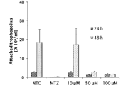
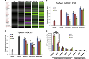
Invitro two-dimensional migration assay
A Oris 96-well plate (Oris migration assaykit) was coated with 9mg/ml rat-tail collagen and incubated for 30–45 minutesat 37oC/5% CO2. After incubation, Oris cell seedingstoppers were inserted according to the manufacturer’s instructions.Serum-starved KFs and ELFs were pre-labeled with PKH26 according to themanufacturer’s instructions. A density of 2.5X10⁵ cells per well wasseeded in each well of the Oris 96-well migration assay plates. The plate wasthen incubated overnight at 37 oC/5% CO2.The next day, the cell seeding stoppers were removed and 100μlof fresh medium was added with or without different compounds as above; theplates were further incubated and the cells were allowed to migrate for B30hours in the migration zone. Micrographs were captured using x4 magnificationof inverted microscopy. Cells in the migration zone were counted from four independentexperiments and average migrated cells were plotted on the graphs.
Invitro three-dimensional invasion assay
Inhibition of the invasive capacity ofKU-0063794, KU-0068650, and Rapamycin was tested using basement membraneextract in vitro in three-dimensional invasion assay (Oris Invasion anddetection assay kit). Briefly, serum-starved cells at a density of 2.5x10⁵ cells per well were seeded in Oris invasion assay plates and allowed to attachfor 8–12 hours at 37 oC/5% CO2; after cell attachment, thestoppers were removed from the wells and cells were washed once withphosphate-buffered saline and 40μl of basement membrane extractwas added to the cells. The plates were incubated (at 37 oC/5% CO2)for 45–60 minutes. Compound treatments were given for 48 hours and cells wereallowed to invade in the 2-mm invasion zone created by Oris cell seedingstoppers. The cells were stained with Calcein AM according to themanufacturer’s instructions. Micrographs were captured using x4 magnification ofinverted Olympus IX71 microscopy. Invaded cells in the invasion zone werecounted from four independent experiments and average invaded cells wereplotted on the graphs.
-
动物实验
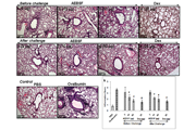
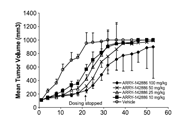
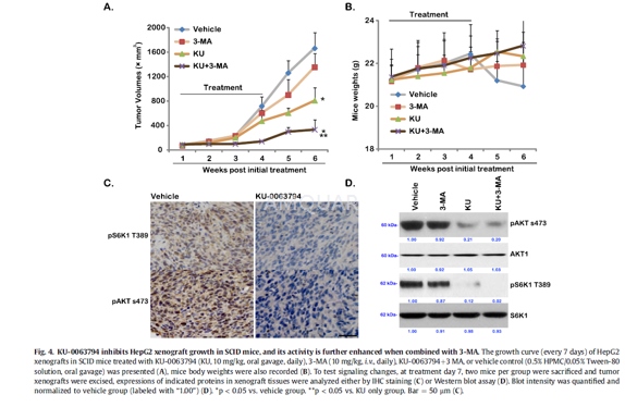
Xenograft model[3]
Eight-week-old female severe combined immunodeficient(SCID)mice were used. Five million of HepG2 cells per mice were injected into theright flanks, and tumors were allowed to reach 10 mm in maximal diameter. Mice(n = 10 each group) were then treated once daily with either vehicle control(0.5% HPMC/0.05% Tween-80 solution, oral gavage), KU-0063794 (10 mg/kg, oralgavage), 3-MA (10 mg/kg, i.v.), or 3-MA + KU-0063794 for 21 days. Mice bodyweights and bi-dimensional tumor measurements were taken every 7 days. Tumorvolumes were estimated using the standard formula: (length x width2)/2.Two mice per group were sacrificed 7 days after initial treatment, and theprimary tumors were excised for analysis. Tumor xenografts were stored inliquid nitrogen.
Immunohistochemistry(IHC)
Paraffin-embedded HepG2 xenograft tissueswere consecutively sectioned at 4μM intervals and mounted onpolylysine-coated glass slides. Slides were incubated at 60oC for1e2 h and then deparaffinized and rehydrated. Antigen retrieval was performedin citrate buffer (pH 6.0) in pressure cooker. Endogenous peroxidase activity wasblocked by incubation with 3% hydrogen peroxide for 15 min at room temperature.Slides were subsequently incubated with primary antibodies againstphosphorylated AKT (p-AKT Ser-473) (1:25 dilution) or phosphorylated S6K1 (1:50dilution) overnight at 4oC. After incubation at room temperature for30 min with biotin-free horseradish peroxidase (HRP) enzyme labeled polymer ofEnVision plus detection system, the slides were developed with the 3,3’- diaminobenzidinesolution followed by counterstaining with hematoxylin.
-
不同实验动物依据体表面积的等效剂量转换表(数据来源于FDA指南)
| 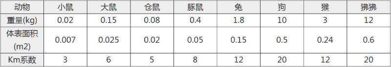 动物 A (mg/kg) = 动物 B (mg/kg)×动物 B的Km系数/动物 A的Km系数 |
|
例如,已知某工具药用于小鼠的剂量为88 mg/kg , 则用于大鼠的剂量换算方法:将88 mg/kg 乘以小鼠的Km系数(3),再除以大鼠的Km系数(6),得到该药物用于大鼠的等效剂量44 mg/kg。
-
参考文献
[1] Garcia-Martinez JM, Moran J, Clarke RG, et al. Ku-0063794 is a specific inhibitor of the mammalian target of rapamycin (mTOR). Biochem J. 2009;421(1):29-42.
[2] Syed F, Sanganee HJ, Singh S, Bahl A, Bayat A. Potent dual inhibitors of TORC1 and TORC2 complexes (KU-0063794 and KU-0068650) demonstrate in vitro and ex vivo anti-keloid scar activity. J Invest Dermatol. 2013;133(5):1340-1350.
[3] Yongxi T, Haijun H, Jiaping Z, Guoliang S, Hongying P. Autophagy inhibition sensitizes KU-0063794-mediated anti-HepG2 hepatocellular carcinoma cell activity in vitro and in vivo. Biochem Biophys Res Commun. 2015;465(3):494-500.
分子式
C25H31N5O4 |
分子量
465.54 |
CAS号
938440-64-3 |
储存方式
﹣20 ℃冷藏长期储存。冰袋运输 |
溶剂(常温)
|
DMSO
15 mg/mL |
Water
<1 mg/mL |
Ethanol
<1 mg/mL |
体内溶解度
-
Clinical Trial Information ( data from http://clinicaltrials.gov )
注:以上所有数据均来自公开文献,并不保证对所有实验均有效,数据仅供参考。
-
相关化合物库
-
使用AMQUAR产品发表文献后请联系我们