-
生物活性
GNF-5837 is a potent pan-Trk inhibitor. GNF-5837 displays antiproliferative effects in cellular Ba/F3 assays (IC50 values are 7, 9 and 11 nM for cells containing the fusion proteins Tel-TrkC, Tel-TrkB and Tel-TrkA, respectively). Exhibits selectivity for Trk receptors over a range of kinases, with some activity at PDGFR and c-Kit (IC50 values are 0.87 and 0.91 μM respectively).
-
体外研究
-
体内研究
-
激酶实验
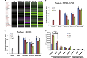
Inhibition of biochemical TrkA, TrkB and TrkC[1]
TrkA and TrkC biochemical assays werecarried out by HTRF method. The reaction mixtures contained 1μM peptidesubstrate, 1μM ATP, and either 1.8nM TrkA or 34nM TrkC in the reaction buffer(50mM HEPES pH 7.1, 10mM MgCl2, 2mM MnCl2, 0.01% BSA,2.5mM DTT and 0.1mM Na3VO4) at a final volume of 10μL.All reactions were carried out at room temperature in white ProxiPlate™384-well Plus plates and were quenched with 5μL of 0.2M EDTA at 60 min. Five μLof the detection reagents (2.5ng PT66K and 0.05μg SAXL per well) were added,the plates were incubated at room temperature for 1 h and then read in EnVisionreader. Compounds were diluted into assay mixture (final DMSO 0.5%), and IC50values were determined by 12-point (from 50 to 0.000282μΜ) inhibition curves induplicate under the assay conditions as described above.
TrkB biochemical assay was carried out bycaliper microfluidic method. The peptide substrate used was FAM-KKKKEEIYFFF-CONH2.The reaction mixtures contained 1μM peptide substrate, 10μM ATP, and 2nM TrkBin a reaction buffer containing 100mM HEPES, pH 7.5, 5mM MgCl2,0.01% Triton X-100, 0.1% BSA, 1mM DTT, 10μM Na3VO4,and 10μM Beta-Glycerophosphate. The reactions were carried out at roomtemperature for 3 hrs, and the products were determined by Caliper EZ-reader.Compounds were diluted into assay mixture (final DMSO 1%), and IC50 values weredetermined by 12-point (from 50 to 0.000282 μM) inhibitioncurves in duplicate under the assay conditions as described above.
-
细胞实验
Cancer cell growth assay[2]
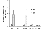
The antiproliferative efficacy of GNF-5837breast cancer cell lines (MCF-7, MDA-MB-231 and SKBR-3) using resazurin assay.Briefly, cells were seeded into 96-well plates in 100-μl media containing FBS10% and incubated for 48 h at 37°C in humidified air with 5% CO2.Cells (70% confluent) were then treated with equivalent concentrations (0–40μM) of free GNF-5837 in media containing FBS 2.5%. Control NPs were also usedfor each concentration. 48-h post-treatment, the media were changed with freshmedia containing 20% v/v resazurin solution for an additional 4 h to assess therelative anticancer activity since only viable cells can reduce nonfluorescentresazurin to highly fluorescent resorufin. The fluorescent intensity of each wellwas then quantitated by Fluostar OPTIMA plate reader (excitation 544 nm andemission 590 nm) and the rate of cell viability was drawn on a dose-responsecurve. The sensitivity to the administered therapies was assessed by the IC50.
Cancercell apoptosis assay
The impact of treatments on the apoptosisprofile of breast cancer cells was evaluated through Hoechst staining. Briefly,cells were grown on coverslips in 24-well plates in media containing FBS 10%.48-h postincubation, cells were treated in triplicates with control mediacontaining FBS 2.5%, 10-μM grade concentrations of free GNF-5837 for another48h. Cells grown on coverslips were then rinsed with ice-cold phosphate-bufferedsaline (PBS), fixed with cold methanol, rinsed again and stained with 1-μg/ mlHoechst 33258 (Life Technologies) in the dark. Stained cells were finallyrinsed and mounted with coverslips using Glycergel on microscope slides.Apoptotic cells exhibiting condensed/ fragmented nuclei were counted under theAxioplan2 upright fluorescence microscope with 4’,6-diamidino-2-phenylindole(DAPI) filter in randomly selected fields (∼1 mm, 12 fields per case at 200× magnification). A minimum of250–400 nuclei were examined for each case and final results were expressed asthe average of the percentage of apoptotic bodies per field.

-
动物实验
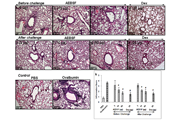
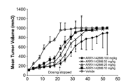
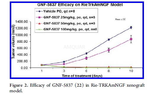
Rie-TRKA/NGF xenograft tumor model[1]
3 x 10⁶ of Rie cells expressingTrkA and NGF were implanted with a subcutaneous injection into the right hindflank of a Balb/c nude mouse. After implant, once tumors became palpable, animalswere dosed with the vehicle, 25 mg/Kg, 50 mg/Kg, or 100 mg/Kg of GNF-5837 usingoral gavages once a day for 10 days. Tumor volumes were measured by a caliper 3times per week and were calculated using (L x W x H)/2.
-
不同实验动物依据体表面积的等效剂量转换表(数据来源于FDA指南)
| 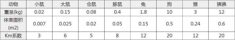 动物 A (mg/kg) = 动物 B (mg/kg)×动物 B的Km系数/动物 A的Km系数 |
|
例如,已知某工具药用于小鼠的剂量为88 mg/kg , 则用于大鼠的剂量换算方法:将88 mg/kg 乘以小鼠的Km系数(3),再除以大鼠的Km系数(6),得到该药物用于大鼠的等效剂量44 mg/kg。
-
参考文献
[1] Albaugh P, Fan Y, Mi Y, et al. Discovery of GNF-5837, a Selective TRK Inhibitor with Efficacy in Rodent Cancer Tumor Models. ACS Med Chem Lett. 2012;3(2):140-145.
[2] Shargh VH HH, Liang M. Gelatin-albumin hybrid nanoparticles as matrix metalloproteinases-degradable delivery systems for breast cancer therapy. Nanomedicine (Lond). 2017;12(9):977-989.
分子式
C28H21F4N5O2 |
分子量
535.49 |
CAS号
1033769-28-6 |
储存方式
﹣20 ℃冷藏长期储存。冰袋运输 |
溶剂(常温)
|
DMSO
100 mM |
Water
<1 mg/mL |
Ethanol
10 mM |
体内溶解度
-
Clinical Trial Information ( data from http://clinicaltrials.gov )
注:以上所有数据均来自公开文献,并不保证对所有实验均有效,数据仅供参考。
-
相关化合物库
-
使用AMQUAR产品发表文献后请联系我们