-
生物活性
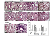
BIO is a cell-permeable, highly potent, selective, reversible, and ATP-competitive inhibitor of glycogen synthase kinase-3α (GSK-3α) and glycogen synthase kinase-3β (GSK-3β; IC50 = 5 nM). GSK-3 plays important roles in numerous signaling pathways that regulate a variety of cellular processes including cell proliferation, differentiation, apoptosis and embryonic development. GSK-3 Inhibitor IX also inhibits phosphoinositide-dependent kinase 1 (PKB Kinase or PDK1), a master kinase responsible for the activation of AKT/PKB, PKC, p70 S6 kinase (S6K), and serum/glucocorticoid regulated kinase (SGK). Research shows that PKB Kinase inhibition halts endothelial cell migration. This small molecule has been been widely used in embryonic stem cell studies to maintain human embryonic stem cells in their undifferentiated state and to sustain expression of the pluripotent state-specific transcription factors Oct-3/4, Rex-1 and Nanog. GSK-3 Inhibitor IX has been shown to induce the differentiation of neonatal cardiomyocytes and maintain self-renewal in both human and mouse embryonic stem cells through Wnt signaling activation. GSK-3 Inhibitor IX is also an inhibitor of Cdk5 and p35. Maintains self-renewal and pluripotency in embryonic stem cells via activation of the Wnt signaling pathway in vitro; also promotes proliferation and dedifferentiation in cardiomyocytes.
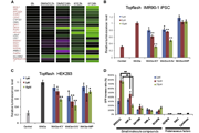
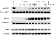
Kinase activities of BIO

AhRactivation of 6BIO[4]

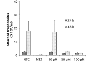
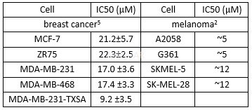
Anti-proliferative effects of BIO
-
体外研究
-
体内研究
30% PEG400+0.5% Tween80+5% propylene glycol
-
激酶实验
Kinase assays[1]
Buffers
Homogenization Buffer - 60 mM β-glycerophosphate,15mM p-nitrophenylphosphate, 25mM Mops (pH 7.2), 15mM EGTA, 15mM MgCl2,1mM DTT, 1mM sodium vanadate, 1mM NaF, 1mM phenylphosphate, 10μg leupeptin/ml,10μg aprotinin/ml, 10μg soybean trypsin inhibitor/ml and 100μM benzamidine.
Buffer A - 10mM MgCl2, 1 mMEGTA, 1 mM DTT, 25mM Tris-HCl pH 7.5, 50μg heparin/ml.
Kinasepreparations and assays
Kinase activities were assayed in Buffer A,at 30°C, at a final ATP concentration of 15μM. Blank values were subtracted andactivities calculated as pmoles of phosphate incorporated during a 10min.incubation. The activities are usually expressed in % of the maximal activity,i.e. in the absence of inhibitors. Controls were performed with appropriatedilutions of dimethylsulfoxide.
GSK-3α/β was purified from porcine brain byaffinity chromatography on immobilized axin. It was assayed, following a 1/100dilution in 1mg BSA/ml 10mM DTT, with 5μl 40μM GS-1 peptide, a specific GSK-3substrate, (YRRAAVPPSPSLSRHSSPHQSpEDEEE), in buffer A, in the presence of 15μM[γ-32P] ATP (3,000Ci/mmol; 1mCi/ml) in a final volume of 30μl. After30min. incubation at 30°C, 25μl aliquots of supernatant were spotted onto 2.5 x3 cm pieces of Whatman P81 phosphocellulose paper, and, 20sec. later, thefilters were washed five times (for at least 5 min. each time) in a solution of10 ml phosphoric acid/liter of water. The wet filters were counted in thepresence of 1 ml ACS scintillation fluid.
-
细胞实验
Cell culture and transfection[6]
The OC cell lines A2780 and OVCAR3 werewere maintained in Roswell Park Memorial Institute (RPMI)-1640 (OVCAR3) orDulbecco’s modified Eagle’s medium (DMEM) media supplemented with 10 % fetalbovine serum (FBS), 100 U/mL penicillin, and 100 μg/ mL streptomycin in ahumidified atmosphere of 5 % CO2 at37 °C. The GSK-3β and mock small interferingRNA (siRNA) used to transfect the A2780 and OVCAR3 cells. To evaluate theeffect of BIO, the dissolved drug was added to A2780 and OVCAR3 at different concentrations(0, 2, 4, and 8 μM).
Proliferationassay
Cell proliferation was analyzed using the3-(4,5-dimethylthiazol-2-yl)-2, 5-diphenyltetrazolium bromide (MTT) assay. Cellswere seeded into 96-well plates (3×10³ cells/well) directly andallowed to adhere. At different time points, 0, 24, 48, and 72 h, 20 μL of MTT(5 mg/mL) was added to cells. Incubation at 37 °C for 4 h was carried out.Supernatants were removed, and 150μL of dimethylsulfoxide (DMSO) was added to eachwell. The absorbance value of each well was measured at490 nm.

-
动物实验
Experimental design[7]
Adult male C57BL/6 mice weighing 24–28 gand in the age range of 8–10weeks old were used in this study.
Mice were randomly divided into ischemicstroke-vehicle group and stroke plus BIO treatment group. Three days afterischemia, BIO 8.5μg/kg or equivalent volume of DMSO was administered intraperitoneallyevery 2 days until sacrificed at 14 or 21 days after stroke. To label proliferatingcells, 5-bromo-20-deoxyuridine (BrdU) (50mg/kg/day, i.p.) was administered toall animals beginning on day 3 after ischemia until the day of sacrifice.
Although BIO is an established inhibitor ofGSK-3β, there is limited information about its effective dosages in vivo.High doses of 50 mg/kg BIO was tested and showed certain cytotoxic effect. BIOwas also given at 0.2 mg/kg for repeated intraperitoneal (i.p.)administrations. Alow dosage of 8.5μg/kg BIO was selected based on itseffect of reducing the downstream molecule phosphorylated GSK-3β.
Westernblot analysis
Fresh brain tissue was isolated under amicroscope from the peri-infarct area defined as the region within 500μmfrom the edge of the infarct area. Western blot analysis was performed toanalyze protein expression in penumbra. In brief, brain tissue was lysed withmodified radioimmunoprecipitation assay buffer (50mmol/L HEPES, pH 7.3, 0.1%sodium deoxy-cholate, 150mmol/L NaCl, 1mmol/L ethylenediaminetertraacetic acid,1mmol/L sodium orthovanadate, 1mmol/L NaF), and protease inhibitor cocktail withcontinuous manual homogenization. After 30min, lysate was spun at 17,000 rpmfor 15 min at 4oC and supernatant was collected. Proteinconcentration was deter-mined using the Bicinchoninic Acid Assay. Equal amountsof protein (30μg) were electrophoresed on 6%–20% sodium dodecylsulfate-polyacrylamide gradient gel (SDS-PAGE) in a Hoefer Mini-Gel system and transferredin the Hoefer Transfer Tank to a polyvineylidene difluride (PVDF) membrane.Membranes were blocked with 5% BSA in Tris bufferedsaline (TBS, 20mmol/L Tris,137mmol/l NaCl and 0.1% tween-20) at room temperature for 1h and incubatedovernight at 4oC with primary antibodies against brain-derivedneurotrophic factor (BDNF) (1:500), GAP-43(1:500), β-catenin(1:1000). β-tubulin (1:2500) was used as the protein loading control. After 3washes with TBST, blots were incubated with alkaline phosphatase-conjugatedanti-mouse or anti-rabbit IgG antibodies for 2 h at room temperature. Finally,membranes were washed with TBST and the signal was developed by the addition of5-bromo-4-chloro-3-indolylphosphate/nitro blue tetrazolium (BCIP/NBT) solution,quantified, and analyzed by the imaging software ImageJ. The level of proteinexpression was normalized to β-tubulin controls.

-
不同实验动物依据体表面积的等效剂量转换表(数据来源于FDA指南)
|  动物 A (mg/kg) = 动物 B (mg/kg)×动物 B的Km系数/动物 A的Km系数 |
|
例如,已知某工具药用于小鼠的剂量为88 mg/kg , 则用于大鼠的剂量换算方法:将88 mg/kg 乘以小鼠的Km系数(3),再除以大鼠的Km系数(6),得到该药物用于大鼠的等效剂量44 mg/kg。
-
参考文献
[1] Meijer L, Skaltsounis A-L, Magiatis P, et al. GSK-3-Selective Inhibitors Derived from Tyrian Purple Indirubins. Chemistry & Biology. 2003;10(12):1255-1266.
[2] Liu L, Nam S, Tian Y, et al. 6-Bromoindirubin-3'-oxime inhibits JAK/STAT3 signaling and induces apoptosis of human melanoma cells. Cancer Res. 2011;71(11):3972-3979.
[3] Tsakiri EN, Gaboriaud-Kolar N, Iliaki KK, et al. The Indirubin Derivative 6-Bromoindirubin-3'-Oxime Activates Proteostatic Modules, Reprograms Cellular Bioenergetic Pathways, and Exerts Antiaging Effects. Antioxid Redox Signal. 2017.
[more]
分子式
C16H10BrN3O2 |
分子量
356.17 |
CAS号
667463-62-9 |
储存方式
﹣20 ℃冷藏长期储存。冰袋运输 |
溶剂(常温)
|
DMSO
>5 mg/mL |
Water
<1 mg/mL |
Ethanol
10 mM |
体内溶解度
约25 mg/mL
-
Clinical Trial Information ( data from http://clinicaltrials.gov )
注:以上所有数据均来自公开文献,并不保证对所有实验均有效,数据仅供参考。
-
相关化合物库
-
使用AMQUAR产品发表文献后请联系我们


