-
生物活性
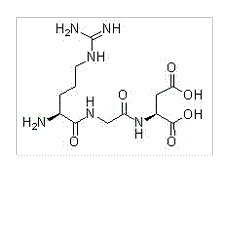
RGD (Arg-Gly-Asp) Peptides is a cell adhesion motif which can mimic cell adhesion proteins and bind to integrins. RGD (Arg-Gly-Asp) Peptides, the cell adhesion peptides, has an affinity for αvβ3 integrinsand has been used to modify carrier-materials for enhancing receptor-mediatedcell adhesion rates, which further contributes to the endocytosis.
-
体外研究
-
体内研究
-
激酶实验
Coating Plates or Latex Beads with Polylysine orFibronectin and Cell Adhesion[1]
Polylysine (10μg/ml) orfibronectin (10μg/ml) in PBS was added to plates (2ml/35-mm plate) and incubated at4 °C overnight. The plates were subsequently washed (3 times) with BSA (2mg/ml)in PBS and then incubated at 37 °C with 2 ml of BSA (2mg/ml) in PBS for1.5–2 h.The plates were then washed (3 times) with PBS and ready for use. For BSAcontrol plates, treatment procedures were the same as described above. Cellsremained in suspension and did not attach to BSA-coated plates. The coating oflatex beads was performed essentially. Briefly, 20 ml of the latex beads were incubatedwith either polylysine (50μg/ml) or fibronectin (50μg/ml) in PBS overnight at 4 °Cwith rotation. After washing with PBS (3 times), the beads were incubated withBSA (2mg/ml) at 37 °C for 2 h, washed with PBS (2 times), resuspended in 200μlof DMEM, and then were ready for use. For cell adhesion, the cells weretrypsinized, resuspended with trypsin inhibitor solution, and washed twice withserum-free DMEM medium. The cells were then resuspended in DMEM. In some cases,the cells were preincubated with DMEM plus cycloheximide or anti-integrinantibodies or RGD peptides at 37 °C for 30 min. The cells were finally addedonto substratum precoated culture dishes or mixed with latex beads andincubated at 37 °C for the indicated time and directly lysed with lysis buffer.
JNKAssays
The flag-tagged JNK1 was immunoprecipitatedwith anti-flag antibody (M5) from the lysates of cells that were transiently transfectedwith pcDNA3 flag-JNK1 or pcDNA3 flag-JNK1 plus pcDNA3 Myc-ACK-2 and/or pcDNA3HA-Cdc42(T17N). The immunocomplex beads were washed twice with lysis buffer andonce with JNK assay buffer (20mM Hepes, pH 7.4, 10mM MgCl2, 1mMsodium orthovanadate) and then were mixed with the kinase assay buffer and GST-Jun(5μg). The phosphorylation was initiated by adding 5μCiof [γ-32P]ATP (17 mCi/nmol) and performed at 22 °C for 20 min.The reaction was stopped by adding 2x SDS sample buffer, and the samples wereboiled for 7 min and loaded onto a 10% SDS-polyacrylamide gel. The gel wastransferred onto a polyvinylidene difluoride membrane that was used forautoradiography and immunoblotting.

-
细胞实验
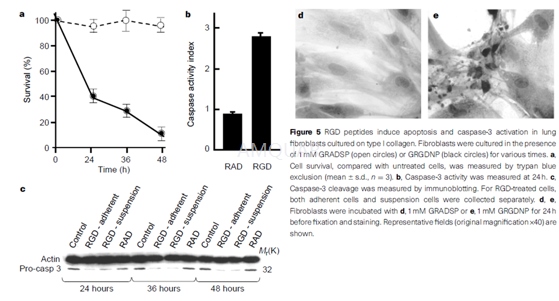
RGD peptides and coated beads[2]
Peptides were added to cell cultures at thestated doses. All peptides were resuspended in PBS and stored in aliquots at-70 oC. All peptides were purified by high-performance liquidchromatography and were .95% pure. Streptavidin-coated Dynabeads (M-280, 2.8μmdiameter, Dynal) were coated with biotinylated peptides according to themanufacturer's instructions.
Detectionof apoptosis and caspase protease activity
Cell death was analysed by the exclusion ofpropidium iodide (5μg/ml) and by forward- and side-scatter characteristics. Results areexpressed as the percentage of live cells remaining compared to the startingcell number. Lung fibroblasts were cultured on bacteriological plastic or glasscoverslips coated with type I collagen (10μg/ml in PBS)for 1h before the addition of RGD-containing peptides. Viable adherent cells wecounted using trypan blue exclusion and samples were taken with which to assesscaspase-3 processing and enzymatic activity. Cells grown on glass coverslipswere fixed in acetone and stained with haemalum to confirm apoptosis. Enzymaticcaspase-1 and caspase-3 activity was measured using the CaspACE fluorometricassay system according to the manufacturer's protocol. Caspase activation isshown as the ratio between the caspase activity of the sample and that measuredin control (non-treated) samples; for all samples, the background activityremaining after inhibition by either caspase-3 (DEVD-CHO) or caspase-1(YVAD-CHO) inhibitors was subtracted.
-
动物实验
-
不同实验动物依据体表面积的等效剂量转换表(数据来源于FDA指南)
|  动物 A (mg/kg) = 动物 B (mg/kg)×动物 B的Km系数/动物 A的Km系数 |
|
例如,已知某工具药用于小鼠的剂量为88 mg/kg , 则用于大鼠的剂量换算方法:将88 mg/kg 乘以小鼠的Km系数(3),再除以大鼠的Km系数(6),得到该药物用于大鼠的等效剂量44 mg/kg。
-
参考文献
[1] Yang W LQ, Guan JL, Cerione RA. Activation of the Cdc42-associated tyrosine kinase-2 (ACK-2) by cell adhesion via integrin beta1. J Biol Chem. 1999;274(13):8524-8530.
[2] Buckley CD PD, Henriquez NV, Parsonage G, Threlfall K, Scheel-Toellner D, Simmons DL, Akbar AN, Lord JM, Salmon M. RGD peptides induce apoptosis by direct caspase-3 activation. Nature. 1999;397(6719):534-539.
分子式
C12H22N6O6 |
分子量
346.34 |
CAS号
99896-85-2 |
储存方式
﹣20 ℃冷藏长期储存。冰袋运输 |
溶剂(常温)
|
DMSO
<1 mg/mL |
Water
10 mg/mL |
Ethanol
<1 mg/mL |
体内溶解度
-
Clinical Trial Information ( data from http://clinicaltrials.gov )
注:以上所有数据均来自公开文献,并不保证对所有实验均有效,数据仅供参考。
-
相关化合物库
-
使用AMQUAR产品发表文献后请联系我们


