-
生物活性
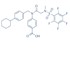
SH-4-54 is a novel, structurally unique small molecule inhibitor thateffectively targets interactions with the pTyr-SH2 domain to block both STAT3and 5 dimerization and subsequent DNA-binding. SH-4-54 is a most potent, small molecule, nonphosphorylated STAT3 inhibitor, strongly binds to STAT3 protein (KD = 300 nM), is selective for STAT3 over STAT1. H-4-54 potently kills glioblastoma brain cancer stem cells (BTSCs) and effectively suppresses STAT3 phosphorylation and its downstream transcriptional targets at low nM concentrations.
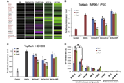
Bindingpotency of SH-4-54[1]

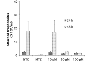
Cytotoxicityagainst GBM BTSC line of SH-4-54[1]

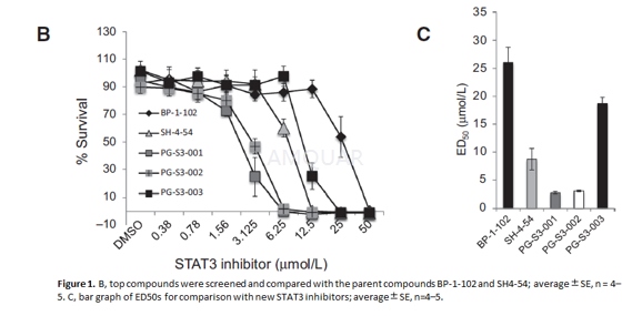
SH-4-54blocks proliferation of Panc10.05 cells with an ED50 value of 8.7±1.9μM.[2]
-
体外研究
-
体内研究
-
激酶实验
Surface Plasmon Resonance (SPR) studies[1]
Interactions of His-tagged STAT3 with smallmolecules were investigated using SPR spectroscopy. The binding experimentswere carried out on a ProteOn XPR36 biosensor at 25°C using the HTE sensorchip. The flow cells of the sensor chip were loaded with a nickel solution at30μL/min for 120s to saturate the Tris–NTA surface with Ni (II) ions. PurifiedHis-tagged STAT3 and STAT5 in PBST buffer (PBS with 0.005% (v/v) Tween-20 and0.001% DMSO pH 7.4) was injected in the first and second channels of the chiprespectively in the vertical direction at a flow rate of 25μg/μL for 300s,which attained, on average, ~8000 resonance unit (RU). After a wash with PBSTbuffer, inhibitor binding to the immobilized proteins was monitored byinjecting a range of concentrations along with a blank at a flow rate of100μL/min for 200s for each of these small molecules. When the injection of thesmall molecule inhibitor was completed, running buffer was allowed to flow overthe immobilized substrates for the non-specifically bound inhibitors todissociate for 600 s. Following dissociation of the inhibitors, the chipsurface was regenerated with an injection of 1M NaCl at a flow rate of 100μL/mlfor 18s. Interspot channel reference was used for non-specific bindingcorrections and the blank channel used with each analyte injection served as adouble reference to correct for possible baseline drift. Data were analyzedusing ProteOn Manager Software version 3.1. The Langmuir 1:1 binding model wasused to determine the KD values.

-
细胞实验
Cell lines and patient-derived PDAC cells[2]
Normal lung fibroblasts, CCD-13Lu, MIA-PaCa-2,Pa03C, Panc10.05, Pa02C and CAF19 were maintained at 37oC in 5% CO2,grown in DMEM with 10% serum, and mycoplasma-free. The CAF19 cells weretransduced with a lentivirus vector to make them stably express enhanced GFP,and Pa03C cells were transduced with a lentivirus vector to make them stablyexpress TdTomato. CAF19 cells were seeded 24 hours before 150pfu/cell of thelentivirus was added to the media. One day later, the virus was removed and thenthe cells were grown for an additional 2 days in regular media.
Survivaland proliferation studies
The proliferative capacity of PDAC and CAF19cells as a monolayer was assessed using MTS tetrazolium dye assay. Using96-well plates, we seeded either tumor cells alone (2,000–3,000 cell/well),CAFs alone (4,000 cell/ well), or tumor + CAFs at a ratio of 2:1. STAT3inhibitors were added 24 hours after the cells were seeded and MTS assay was performed72 hours later.
-
动物实验
Cell Lines, Culture, and Production ofSH-4-54-Resistant Cell Lines[3]
For clonal selection, MDA-MB-231 humanbreast cancer cells lines were plated at several different densities into 10-cmdishes in either DMEM or RPMI supplemented with 10% fetal bovine serum tosupport the optimal growth of MDA-MB-231. Media was changed every 2–3 days toadminister SH-4-54 from a freshly thawed aliquot. After one or two months ofcontinuous drug selection for MDA-MB-231 cells, individual clones were isolatedby “picking” them using sterile cloning discs presoaked in trypsin-EDTA. Eachindividual clone was transferred into one well of a 48-well plate and culturedto confluence in the presence of SH-4-54 prior to reseeding into a larger well format.For experiments, cells were plated into 6-well tissue culture-treated plates at2.5x105 cells/well 24 hours prior to manipulation. Untreatedparental MDA-MB-231 cells, referred to as wild-type counterparts, were passagedin parallel. All cells were determined to be mycoplasma-free.
Drugs
SH-4-54 was reconstituted in DMSO at a 25mMstock. Individual aliquots were stored at -20°C, and cells were treated withvehicle or an appropriate concentration of drug (initially at 10μM, followed bya 5μM maintenance dose).
TumourXenografts
4–6 week old female immunocompromisedBALB/c nu/nu mice were implanted subcutaneously with 21-day release, 0.25 mgpellets of 17β-estradiol three days prior to subcutaneous injection with eitherMDA-MB-231 wild-type or MDA-MB-231 clone #2 (SH-4-54-resistant) cells preparedin sterile phosphate-buffered saline at 4×10⁶ cells in 100μL peranimal. The viability of cells to be injected was assessed via trypan blue exclusionstaining. Pellet implants are routinely carried out to promote the growth ofMDAMB-231 cells in our xenograft models. Two animals were included for eachgroup. Tumour size was evaluated every 3 days, and animals were sacrificed onDay 25 post-injection, when tumours in the wild-type group reached a meanvolume of 794 mm3. Tumours were excised, flash-frozen in liquid nitrogen, andstored at -80°C.

-
不同实验动物依据体表面积的等效剂量转换表(数据来源于FDA指南)
|  动物 A (mg/kg) = 动物 B (mg/kg)×动物 B的Km系数/动物 A的Km系数 |
|
例如,已知某工具药用于小鼠的剂量为88 mg/kg , 则用于大鼠的剂量换算方法:将88 mg/kg 乘以小鼠的Km系数(3),再除以大鼠的Km系数(6),得到该药物用于大鼠的等效剂量44 mg/kg。
-
参考文献
[1] Haftchenary S, Luchman HA, Jouk AO, et al. Potent Targeting of the STAT3 Protein in Brain Cancer Stem Cells: A Promising Route for Treating Glioblastoma. ACS Med Chem Lett. 2013;4(11):1102-1107.
[more]
分子式
C29H27F5N2O5S |
分子量
610.59 |
CAS号
1456632-40-8 |
储存方式
﹣20 ℃冷藏长期储存。冰袋运输 |
溶剂(常温)
|
DMSO
150 mM |
Water
<1 mg/mL |
Ethanol
80 mM |
体内溶解度
-
Clinical Trial Information ( data from http://clinicaltrials.gov )
注:以上所有数据均来自公开文献,并不保证对所有实验均有效,数据仅供参考。
-
相关化合物库
-
使用AMQUAR产品发表文献后请联系我们


