-
生物活性
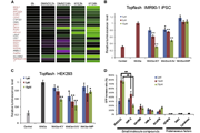
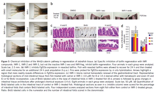
IWR-1-endo is a Wnt signaling protein, which are small secreted proteins that are active in embryonic development, tissue homeostasis, and tumorigenesis. Wnt proteins bind to receptors on the cell surface, initiating a signaling cascade that leads to β-catenin activation of gene transcription. IWR-1-endo is a potent inhibitor of the Wnt response, blocking a cell-based Wnt/β-catenin pathway reporter response with an IC50 value of 180 nM. It inhibits, at 10 μM, Wnt-induced accumulation of β-catenin, leading to proteasomal degradation of this protein through a destruction complex which consists of Apc, Axin2, Ck1, and Gsk3β. IWR-1-endo stabilizes the destruction complex, increasing the level of Axin2 protein without changing the levels of Apc or Gsk3β. The IWR compound does not change the de novo synthesis of Axin2, alter the affinity of Axin2 for β-catenin, or inhibit the proteasome. It has a half-life of 60 minutes in murine whole blood and 20 minutes in intact murine hepatocytes. In in vivo tests, IWR-1-endo inhibits zebrafish tail fin regeneration with a minimum inhibitory concentration of 0.5 μM. The IWR-1-exo diastereomer exhibits much less activity against the Wnt/β-catenin pathway and has been used as a control. IWR-1-endo is an inhibitor of Tankyrase-1 and Tankyrase-2.
Inhibitionof Wnt signaling[1][2]

-
体外研究
-
体内研究
-
激酶实验
-
细胞实验
Cellculture[3]
Skin tissues were washed withphosphate-buffered saline (PBS) supplemented with 1% penicillin-streptomycin.The skin tissues were cut into small pieces, and then placed in culture dish.Fibroblastswere routinely sprouting from explanted tissues in a week, andcultured in Dulbecco's Modified Eagle's Medium (DMEM) supplemented with 10%fetal bovine serum (FBS) and 1% penicillin-streptomycin. The cells passage between3 and 7 were used for the experiments. Before treatment with IWR-1,fibroblastswere incubated in serum-free DMEM overnight.
Cytotoxicityand cell proliferation tests
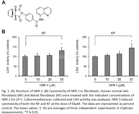
Cytotoxicity was determined by lactatedehydrogenase (LDH) assay. Cells were seeded in 24-well culture plate at adensity of 2 × 10⁴/well, and then treated with differentconcentrations of IWR-1. Culture medium was collected 24 h after treatment with IWR-1 and LDH activity was determined using Cytotoxicity detection kit.
Cell proliferation was measured by cellcounting. The same number of NF or KF was seeded in 60-mm dish. After 24 h,cells were treated with different concentrations of IWR-1. At the indicatedtime points, cells were detached from the culture dish and cell number wascounted using a hemocytometer.
-
动物实验
Zebrafishstudies[2]
Six-month-old zebrafish were incubated 8 or14 d at 28.5 ℃ in aquarium water supplemented with 10μMIWR-1 or in 0.1% (v/v) DMSO as a control. Fish were fed standard diet, andsolutions were changed daily. For BrdU labeling studies, zebrafish wereincubated in 1 mM BrdU in aquarium water for 2 h at room temperature at the endof an 8-d chemical exposure period, then washed several times in aquariumwater, anesthetized with 0.1%(v/v) Tricaine and fixed in 4% (v/v)paraformaldehyde for 48 h at 4 ℃. The intestine was dissected out,dehydrated, paraffin embedded and sectioned at 5-mm intervals. Sections werestained with Hematoxylin and Eosin or processed for BrdU immunohistochemistry.The total number of crypts and total number of BrdU-positive nuclei in the midand distal sections of the intestine were counted from each section. For caudalfin regeneration assays, zebrafish, 3–6 months of age, were anaesthetized in 0.02%(v/v) Tricaine, and half of the fin was resected using a razor blade to remove.Amputees were reared at 28oC in tanks containing either DMSO or IWR(10μM). Water and compounds were replenished daily for the total indicatedassay period. For recovery experiments, fish were then bred in a chemical-freeenvironment at 28oC for an additional 9 d. A 494-base-pair fragment of the Fgf20a codingsequence was amplified with primers AGATGGGACGTCCAGAGATG (forward) andGTCCATGCCAGTGTTTTGTG (reverse) and subcloned into pGEM-T Easy. The resultantplasmid was linearized and an antisense riboprobe was synthesized usingDigoxigenin UTP labeling mix and T7 RNA polymerase according to themanufacturer’s instructions. In situ hybridization of fixed tails was carriedout, except that proteinase K digestion was carried out for 20 min at roomtemperature in 20μg ml–1 proteinase K.
-
不同实验动物依据体表面积的等效剂量转换表(数据来源于FDA指南)
|  动物 A (mg/kg) = 动物 B (mg/kg)×动物 B的Km系数/动物 A的Km系数 |
|
例如,已知某工具药用于小鼠的剂量为88 mg/kg , 则用于大鼠的剂量换算方法:将88 mg/kg 乘以小鼠的Km系数(3),再除以大鼠的Km系数(6),得到该药物用于大鼠的等效剂量44 mg/kg。
-
参考文献
[1] Lu J, Ma Z, Hsieh JC, et al. Structure-activity relationship studies of small-molecule inhibitors of Wnt response. Bioorg Med Chem Lett. 2009;19(14):3825-3827.
[more]
分子式
C25H19N3O3 |
分子量
409.44 |
CAS号
1127442-82-3 |
储存方式
﹣20 ℃冷藏长期储存。冰袋运输 |
溶剂(常温)
|
DMSO
28 mg/mL |
Water
<1 mg/mL |
Ethanol
<1 mg/mL |
体内溶解度
-
Clinical Trial Information ( data from http://clinicaltrials.gov )
注:以上所有数据均来自公开文献,并不保证对所有实验均有效,数据仅供参考。
-
相关化合物库
-
使用AMQUAR产品发表文献后请联系我们


