-
生物活性
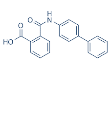
Potently induces differentiation of human mesenchymal stem cells into chondrocytes (EC50 = 100 nM). Kartogenin induces the selective differentiation of multipotent mesenchymal stem cells (MSCs) into chondrocytes. Kartogenin binds to filamin A, and disrupts the specific interaction between filamin A and CBFβ (core-binding factor β subunit). Apparently, kartogenin induces chondrogenesis by regulating the nuclear localization of CBFβ. Reduces disease severity in a mouse model of osteoarthritis; displays protective effects against osteoarthritic stimuli in mature chondrocytes in vitro.
Kartogeninpromotes chondrocyte differentiation from hMSCs in a dose-dependent manner withan EC50 of 100 nM.[1]
-
体外研究
-
体内研究
-
激酶实验
-
细胞实验
BMSCand patellar tendon stem/progenitor cells (PTSC) isolation and culture[2]
BMSCs isolated from the femoral marrowcompartments were cultured in Dulbecco’s modified Eagle’s medium supplementedwith 20% fetal bovine serum100 U/mL penicillin and 100μg/mLstreptomycin in an incubator at 37oC with 5% CO2. Afteran overnight culture, non-adherent cells were removed carefully and replacedwith fresh medium. When primary cultures became almost confluent, cells were treatedwith 0.25% trypsin containing 0.02% ethylenediaminetetraacetic acid for 2 minat room temperature (21oC) and seeded into new culture flasks. Apure population of BMSCs was obtained 3 weeks after the first culture was initiated.
For PTSC isolation, tendon sheaths from thepatellar tendons were removed to obtain the core portions of the tendons.Tendon samples were then minced into small pieces and each 100 mg wet tissuesample was digested in 1 mL phosphate buffered saline (PBS) containing 3 mg collagenasetype I and 4 mg dispase at 37oC for 1 h. After centrifugation at 3500 r/min for 15 min to remove the enzymes, the cells were cultured in growth mediumsupplemented with 100mmol/L 2-mercaptoethanol at 37oC with 5%CO2.The medium was changed every 3 days and cells were split when 90% confluence wasreached. In all further culture experiments, BMSCs and PTSCs at passage 1 or 2were used.
In vitro experiments
Rabbit BMSCs or PTSCs at passage 1–2 wereseeded in 6-well plates at a density of 6x10⁴per well and culturedfor two weeks in growth medium (Dulbecco’s modified Eagle’s medium plus 20%fetal bovine serum) with various concentrations (1 nmol/L–5μmol/L)of KGN. The medium was changed every 3 days and after 2 weeks, cell proliferationwas measured by population doubling time (PDT).
Determinationof PDT for measuring cell proliferation in vitro
Proliferation of KGN treated BMSCs andPTSCs was determined by the time taken for the cells to multiply and was expressedas PDT, which was calculated from the formula PDT=log2[Nc/N0],where N0 is the total number of cells seeded initially and Nc is the totalnumber of cells at confluence.

-
动物实验
Animalsand Study Design[3]
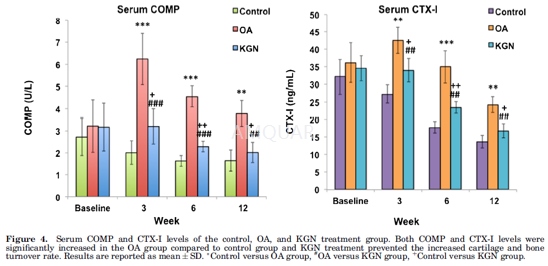
Male Sprague Dawley rats at 4 months of agewere kept in a sanitary ventilated animal room (12-h light/dark cycle) withfood and water available ad libitum. The rats were randomized into threegroups: OA (n=6), KGN treatment (KGN, n=6), and control (n=6) group. On day 0,all the rats from OA and KGN group were anaesthetized with 2% isoflurane, amedial arthrotomy was performed and the anterior cruciate ligament wastransected (ACLT) on the right knee. In the control group rats, only a medialarthrotomy was performed on the right knee, but the intra-articular capsule wasleft intact (sham). Postoperatively, 0.03mg/kg buprenorphine hydrochloride was administeredby subcutaneous injection and as needed subsequently for pain control.
KartogeninTreatment
A stock solution of KGN was prepared bydissolving KGN in dimethyl sulfoxide (DMSO), and then diluted with sterilesaline to prepare the working solution. Rats from KGN treatment group received weeklyintra-articular injections of 125μM KGN in 50μlof saline in the ACL transected knee joint. Rats from the OA group received 50μlof saline in the ACL transected knee joint and the control group rats did not receiveany treatment. The KGN stock solution was diluted104fold withsterile saline to prepare the working solution. This high dilution factoreliminates the potential effects of DMSO on the knee joint if any.
SerumCOMP and CTX-I Analysis
Blood specimens from tail vein wereobtained from all the rats at baseline, 3, 6, and 12 weeks after OA inductionfor measuring serum levels of cartilage oligomeric matrix protein (COMP, acartilage turnover marker), and C-terminal telopeptide of collagen type I(CTX-I, a bone turnover marker). Serum was extracted and frozen in aliquots at-80°Cuntil analyzed. Assays for serum COMP, and CTX-I were performed in triplicatesaccording to the manufacturers’ instructions. The coefficients of variationsfor inter-assay and intra-assay measurements were <16% and <10% for CTX-Iand COMP, respectively.
-
不同实验动物依据体表面积的等效剂量转换表(数据来源于FDA指南)
|  动物 A (mg/kg) = 动物 B (mg/kg)×动物 B的Km系数/动物 A的Km系数 |
|
例如,已知某工具药用于小鼠的剂量为88 mg/kg , 则用于大鼠的剂量换算方法:将88 mg/kg 乘以小鼠的Km系数(3),再除以大鼠的Km系数(6),得到该药物用于大鼠的等效剂量44 mg/kg。
-
参考文献
[1] Johnson K ZS, Tremblay MS, Payette JN, Wang J, Bouchez LC, Meeusen S, Althage A, Cho CY, Wu X, Schultz PG. A stem cell-based approach to cartilage repair. Science. . 2012;336(6082):717-721.
[more]
分子式
C20H15NO3 |
分子量
317.34 |
CAS号
4727-31-5 |
储存方式
﹣20 ℃冷藏长期储存。冰袋运输 |
溶剂(常温)
|
DMSO
70 mg/mL |
Water
<1 mg/mL |
Ethanol
20 mg/mL |
体内溶解度
-
Clinical Trial Information ( data from http://clinicaltrials.gov )
注:以上所有数据均来自公开文献,并不保证对所有实验均有效,数据仅供参考。
-
相关化合物库
-
使用AMQUAR产品发表文献后请联系我们


