-
生物活性
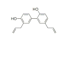
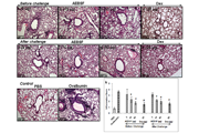
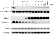
Honokiol is a biphyl neolignan present in the cones, bark, and leaves of Magnolia grandifloris that displays anti-angiogenic, anti-tumor and anxiolytic properties. It inhibits phosphorylation of Akt, p44/42 MAPK, and src. Additionally, honokiol modulates the NF-κB activation pathway, an upstream effector of VEGF, COX-2, and MCL1, all significant pro-angiogenic and survival factors. Honokiol is known to display anti-tumor, anti-angiogenic and anxiolytic properties, and has been shown to induce apoptosis and preferentially inhibit human endothelial cell growth over fibroblasts by VEGFR2 autophosphorylation blocking and subsequent rac activation.
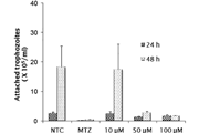
Cellviability ofhonokiol

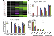
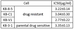
Theanti-proliferation of honokiol in MDR cell lines[5]
Theantioxidant effects of honokiol[6]

-
体外研究
-
体内研究
-
激酶实验
LipidKinase Assays[7]
5μgeach of hemagglutinin-p85 and Myc-p110 were cotransfected into human embryonickidney 293 cells. After 24 h, cells were treated with 10μMhonokiol or same volume of Me2SO as control for another 24 h. Afterremoval of the culture medium, cells were washed with 5 ml of ice-cold PBStwice, lysed in 0.5 ml of lysis Buffer A (50 mM Tris, pH 7.4, 40 mM NaCl, 1 mMEDTA, 0.5% Triton X-100, 1.5 mM Na3VO4, 50mM NaF, 10 mM sodium pyrophosphate,10 mM sodiumβ-glycerophosphate, 1 mM phenylmethylsulfonyl fluoride, 5 mg/mlaprotinin, 1 mg/ml leupeptin, and 1 mg/ml pepstatin A) and was centrifuged for10 min at 14,000μg at 4 °C. P110 was immunoprecipitated with anti-Myc antibody from500μl of the supernatant. The immunoprecipitate was washed with thefollowing buffers: three times with Buffer B (PBS, 1% Nonidet P-40, and 1 mMdithiothreitol); twice with Buffer C (PBS, 0.5 M LiCl, and 1 mMdithiothreitol); and twice with Buffer D (10 mM Tris- HCl, pH 7.4, 0.1 M NaCl,and 1 mM dithiothreitol). After washing, samples were aspirated completely andresuspended in 100μl of kinase buffer (40 mM Tris-HCl, 150 mM NaCl, 20 mM MgCl2, and 1mM dithiothreitol). 10μl of propidium iodide substrate (2 mg/ml in HEPES, pH 7.6, 1 mMEDTA, and 0.1% cholate) was added, and samples were incubated at roomtemperature for 10 min. Reactions were initiated by adding 30 ml of reactionbuffer (70μM ATP in kinase buffer with 10μCi of [γ-32P]ATP/reaction)to each sample. After 10-min incubation at room temperature, the mixturesolubilized in 8μl of 37% HCl was added and vortexed for a few seconds. 150μlof 1:1 CHCl3:CH3OH was introduced and mixed and then centrifuged for 5 min. Thebottom organic layer was removed into a fresh tube and air-dried overnight. Thenext morning 10μl of methanol was added to dissolve the lipid, and then it wasspotted onto a TLC plate and the lipids were separated by 65:35 (v/v)2-propanol, 2 M acetic acid. After the TLC plate was dried, it was exposed to afilm.
-
细胞实验
Celllines[3]
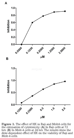
The Raji and Molt4 cell lines were grown at37°C in 5% CO2 in RPMI 1640 medium, with 10% heat-inactivated FBS, 100 units/mLpenicillin and 100 μg/mL streptomycin.
Cellviability assay
Honokiolcytotoxicity was determined by using the colorimetric MTT assay.For this, the cells (1×10⁵ cells/well) were seeded into 96-well platescontaining 100μl of the growth medium, in the absence or presence of increasingconcentrations of HK, at 37°C in 5% CO2for 24 and 48 hrs. MTT (50μl/well, 5 mg/ml in PBS) was then added and incubated for 4 hrs at 37°C. Todissolve the dark blue crystals of formazan 100 μl of DMSO were added. Later,the absorbance was measured in a microplate reader. The experiments wereperformed in triplicate. While the cytotoxicity was determined by plotting, thenumber of affected cells vs drug concentration and inhibition of growth wasexpressed as a percentage of the untreated controls. Thus, the IC50 wasdetermined as the concentration that inhibited cell growth by 50%.
-
动物实验
Invivo Anti-tumor Efficacy Assay[5]
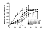
KB-8-5 cells (5x10⁵) wereinjected s.c. into 4–5 week-old female nude mice (Athymic nu/nu). When thetumors had developed to about 100 mm3, the mice were divided intofour groups (n= 7 or 8) in a way to minimize weight and tumor size differencesamong the groups: control group treated with 20% intralipid, honokiol group(1.0 mg/mouse or 50 mg/kg), paclitaxel group (20 mg/kg), and honokiol (1.0 mg/ mouseor 50 mg/kg) plus paclitaxel (20 mg/kg) combination group. Honokiol wasdissolved in 100% ethanol and mixed with 20% intralipid in a 1:14 (v/v) ratio.Honokiol was administered to the mice 3 times per week at 1.0 mg/mouse (or 50mg/kg) via intraperitoneal injection. Paclitaxel was administered to the mice onceper week at 20 mg/kg through tail vein injection. The therapy was continued for4 weeks. The body weight and tumor size were measured three times per week. Thetumor volume was calculated using the formula: V =1/6xlarger diameter x (smallerdiameter).

-
不同实验动物依据体表面积的等效剂量转换表(数据来源于FDA指南)
|  动物 A (mg/kg) = 动物 B (mg/kg)×动物 B的Km系数/动物 A的Km系数 |
|
例如,已知某工具药用于小鼠的剂量为88 mg/kg , 则用于大鼠的剂量换算方法:将88 mg/kg 乘以小鼠的Km系数(3),再除以大鼠的Km系数(6),得到该药物用于大鼠的等效剂量44 mg/kg。
-
参考文献
[1] Sánchez-Peris M, Murga J, Falomir E, Carda M, Marco JA. Synthesis of honokiol analogues and evaluation of their modulating action on VEGF protein secretion and telomerase-related gene expressions. Chemical Biology & Drug Design. 2017;89(4):577-584.
[2] Lai IC, Shih PH, Yao CJ, et al. Elimination of cancer stem-like cells and potentiation of temozolomide sensitivity by Honokiol in glioblastoma multiforme cells. PLoS One. 2015;10(3):e0114830.
[more]
分子式
C18H18O2 |
分子量
266.33 |
CAS号
35354-74-6 |
储存方式
﹣20 ℃冷藏长期储存。冰袋运输 |
溶剂(常温)
|
DMSO
42 mg/mL |
Water
|
Ethanol
<1 mg/mL |
体内溶解度
-
Clinical Trial Information ( data from http://clinicaltrials.gov )
注:以上所有数据均来自公开文献,并不保证对所有实验均有效,数据仅供参考。
-
相关化合物库
-
使用AMQUAR产品发表文献后请联系我们


