-
生物活性
TGX-221 is a potent and selective PI 3-kinase β inhibitor (IC50 values are 0.007, 0.1, 3.5 and 5 μM for the β, δ, γ and α isoforms, respectively). Shows >1,000-fold selectively for PI 3-kinase β over a range of other kinases. Inhibits thrombus formation in animal models. PI 3-Kβ Inhibitor VI, TGX-221 is an excellent tool for studying p110b-dependent responses in cells in vitro.
Lipid kinaseactivity[1]
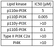
The different inhibition of TGX-221 stereoisomers for p110β-PI3K[2]

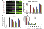
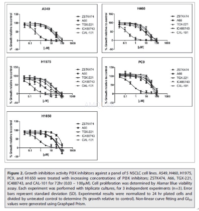
Anti-proliferations of NSCLC cell line[3]

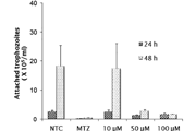
Anti-proliferations of cancer cell lines

-
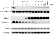
体外研究
-
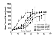
体内研究
1% DMSO+30% polyethylene glycol+1% Tween 80
-
激酶实验
PI3K Lipid Kinase Assays[1][5]
PI3K lipid kinase assays were performed in a total volume of 50 μl in a buffer containing 50 mM HEPES, pH 7.4, 100 mMNaCl, 1 mM dithiothreitol, 5 mMMgCl2, and 100 μM ATP (plus 0.1 μCi of [γ-32P]ATP/assay) using 200 μg/ml phosphatidylinositol as a substrate. Reactions were incubated at 25 °C for 20 min and terminated by the addition of 100 μl of 0.1M HCl and 200 μl of chloroform: methanol (1:1). The mixture was vortexed, and the phases were separated by centrifugation at 10,000 × g for 2 min. The aqueous phase was discarded, and the lower organic phase was washed with 80 μl of methanol:1 M HCl (1:1). After centrifugation, the aqueous phase was again discarded, and the lower organic phase was evaporated to dryness. Reaction products were resuspended in 30 μl of chloroform: methanol (4:1) and spotted onto thin layer Silica Gel 60 plates pretreated with 1% oxalic acid and 1 mM EDTA in water: methanol (6:4). TLC plates were developed in chloroform: methanol: 4 M ammonia (9:7:4). Images of radiolabeled protein and lipid products were analyzed using a Fuji FLA-2000 phosphorimager and Fuji Image Gauge software.
-
细胞实验
Cell lines and culture conditions[3]
Human NSCLC cell lines contain A549, H460, H1650, H1975, and PC9 were obtained. The lines were frozen at low passage for future use and subsequently were confirmed by STR testing. All cell lines were propagated as monolayer cultures at 37oC in 5% CO2using RPMI 1640 growth media supplemented with 10% fetal bovine serum (FBS), glucose, sodium pyruvate, HEPES buffer and penicillin-streptomycin.
Cell proliferation assessment
The growth inhibitory activity (GI) of each compound was tested on cells using the Alamar Blue viability and Trypan Blue exclusion assays. In the Alamar Blue assay, 2x 10³cells/well were plated in 96-well cell culture plates. Cells were treated 24hr later with PI3K inhibitors for 72hr in RPMI 1640 containing 1% serum. For the Trypan Blue exclusion assay, 1x10⁴cells/well were plated in 24-well cell culture plate. The cells were synchronized 24 hr later by changing media to RPMI containing 0.1% serum. After 24hr serum deprivation, cells were released in RPMI containing 1% serum with PI3K inhibitors for 72 hr. Cells were trypsinized, and mixed 1:1 with trypan blue for visual counting of both viable and dead cells. Experimental concentrations from (0.3 – 30μM) of A66, TGX-221, IC488743 and CAL-101 were tested. GI50 values were calculated using Graphpad Prism software.
Cell cycle and apoptosis analysis
For the cell cycle study, NSCLC cell lines were plated in 60- mm dishes. After 24hr, the cells were treated with ZSTK474 and IC488743 at the indicated GI75 concentrations for 24 and 48 hr. Following incubation, cells were trypsinized, quenched with serum, washed, resuspended in phosphate buffered saline (PBS), and fixed in cold 70% ethanol. Cell cycle distribution was assessed by propidium iodide (PI) staining then quantified by BD FACS Calibur flow cytometery or Life Technologies Attune flow cytometry. The data were analyzed with CellQuest software or FlowJo, respectively. An analysis of apoptosis was performed using the same conditions as the cell cycle study, but whole-cell lysates were analyzed by western blot analysis for cleavage of PARP.
-
动物实验
Tail bleeding studies inrats[1]
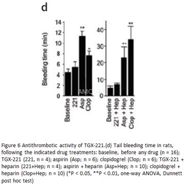
Tail bleeding time was measured in anesthetized rats (halothane in O2) 15 min before and 5 and 30 min after drug administration. We administered the following drug concentrations: TGX-221 (2 mg/kg intravenous), aspirin (200 mg/kg orally), clopidogrel (10 mg/kg orally) and heparin (100 U/kg intravenous). We administered clopidogrel orally 25 h before tail bleeding. Aspirin was administered twice orally: the first dose 25 h before the experiment and the second dose 1 h before the start of the experiment. For experiments involving pretreatment with aspirin or clopidogrel or both, tail bleeding time was also measured before the first gavage dose at 25 h before tail bleeding. Incisions 5 mm long and 1 mm deep47 were made in the tail at each time point and bleeding was monitored by blotting with filter paper48 every 30 sec until it had ceased (tail bleeding time).
-
不同实验动物依据体表面积的等效剂量转换表(数据来源于FDA指南)
|  动物 A (mg/kg) = 动物 B (mg/kg)×动物 B的Km系数/动物 A的Km系数 |
|
例如,已知某工具药用于小鼠的剂量为88 mg/kg , 则用于大鼠的剂量换算方法:将88 mg/kg 乘以小鼠的Km系数(3),再除以大鼠的Km系数(6),得到该药物用于大鼠的等效剂量44 mg/kg。
-
参考文献
[1] Jackson SP, Schoenwaelder SM, Goncalves I, et al. PI 3-kinase p110beta: a new target for antithrombotic therapy. Nat Med. 2005;11(5):507-514.
[2] Lin H, Erhard K, Hardwicke MA, et al. Synthesis and structure-activity relationships of imidazo[1,2-a]pyrimidin-5(1H)-ones as a novel series of beta isoform selective phosphatidylinositol 3-kinase inhibitors. Bioorg Med Chem Lett. 2012;22(6):2230-2234.
more
分子式
C21H24N4O2 |
分子量
364.44 |
CAS号
663619-89-4 |
储存方式
﹣20 ℃冷藏长期储存。冰袋运输 |
溶剂(常温)
|
DMSO
40 mM |
Water
<1 mg/mL |
Ethanol
<1 mg/mL |
体内溶解度
约27 mg/mL
-
Clinical Trial Information ( data from http://clinicaltrials.gov )
注:以上所有数据均来自公开文献,并不保证对所有实验均有效,数据仅供参考。
-
相关化合物库
-
使用AMQUAR产品发表文献后请联系我们


