-
生物活性
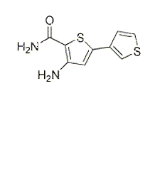
SC-514 is ATP-competitive IKKβ inhibitor (IC50 = 3 - 12 μM) that displays > 10-fold selectivity over 28 other kinases including JNK, p38, MK2 and ERK. Attenuates NF-κB-induced gene expression of IL-6, IL-8 and COX-2 in synovial fibroblasts (IC50 values are 20, 20 and 8 μM respectively).
SC-514inhibits RANKL-induced activation of NF-κB with an IC50 of 2.5μM.[1] SC-514 inhibits various forms of recombinant human IKK-2 with IC50 values of 3–12 μM.[2]
The inhibition of recombinant human IκB kinase (IKK)[2]

The inhibition of tyrosine and serine-threonine kinases[2]

-
体外研究
-
体内研究
-
激酶实验
Kinase Assay[2]
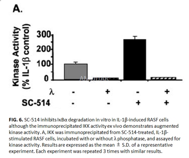
IKK complexes were immunoprecipitated from IL-1β-treated RASF cell lysates (0.5–2 mg) using a NEMO antibody (3–10μg) followed by the addition of protein A-agarose beads. Antibody complexes were pelleted by centrifugation and washed 3 times with 1 ml of cold whole-cell lysis buffer followed by 2 washes in kinase buffer (25mM HEPES, pH 7.6, 2mM MgCl2, 2mM MnCl2, 10mM NaF, 5mM DTT, and 1mM phenylmethylsulfonyl fluoride). 100–200μg of immunoprecipitated IKK was analyzed for kinase activity in a reaction containing 10μM biotinylated IκBαpeptide as substrate and 1μM [γ-33P] ATP (2500 Ci/mmol). After incubation at room temperature for 30 min, 25μl of the reaction mixture was withdrawn and added to a SAMTM 96 biotin capture plate. After successive wash steps the plate was allowed to air-dry and 25μl of scintillation fluid was added to each well. Incorporation of [γ-33P] ATP was measured using a Top-Count NXT. Km determination of rhIKK-2 is as follows. Briefly, various concentrations of [γ-33P] ATP or peptide substrates were used in the assay at a fixed (3xKm) concentration of the second substrate and 100ng of rhIKK-2 in a final volume of 50μl. Following incubation for 30 min at 25 °C, the reaction was stopped by the addition of 150μl of AG1XB resin in 900 mM sodium formate buffer, pH 3 (the resin is in slurry of 1 volume resin to 2 volume of sodium formate buffer). The resin was allowed to settle, and 50μl of supernatant was transferred to a top count plate followed by the addition of 150μl of Microscint 40, mixed well, and counted. For p65 FL-GST, 0.043–2.72μM concentrations were used. Once Km graphs were fitted using GrafitTM program, Kcat values were calculated from Vmax values and expressed as units/mol of enzyme/h.
-
细胞实验
Apoptosis assay[1]
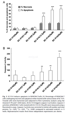
1 x 10⁶RAW264.7 cells were seeded in 2 mL of complete media in a 6-well cell culture plate and then incubated in a humidified atmosphere of 5% CO2and 95% air at 37oC overnight. Media was removed after overnight incubation. SC-514 with various concentrations of 0μM, 3.1μM, 6.2μM, 12.5μM, 25μM, or 50μM was added to the wells, and cells were incubated for 24 h. Cells were collected and resuspended in 0.5 mL of 1xBinding Buffer (BD-Pharmingen, NSW, Australia). Aliquots of cell suspensions (100 mL) were then stained with Annexin V-PE (5 mL: BD-Pharmingen) and/or 7-aminoactino-mycin D (7-AAD) (5 mL: BD-Pharmingen). Cells were incubated in the dark at room temperature for 15 min, followed by addition of 400 mL of 1xBinding Buffer and analysis by flow cytometry. The percentage of apoptotic cells in the population was obtained.
Caspase-3 assay
RAW264.7 cells were seeded (2x10⁶cells/well) into 24-well plates, cultured overnight, and then treated with various doses of SC-514 for 24 h. The treated cells were harvested by trypsinization and vigorous pipetting. The cells were pelleted, washed once in PBS and then lysed by three freeze thaw cycles in 20μL of lysis buffer containing 50 mM HEPES (pH 7.4), 100 mM NaCl, 0.1% CHAPS, 0.1mM EDTA, 1mM DTT, 0.5mM PMSF, 5μg/mL pepstatin A and 10 mg/mL leupeptin. The activity of caspase-3 in lysates was determined using a kinetic assay, in buffer containing 50mM HEPES (pH 7.4), 100mM NaCl, 0.1% CHAPS, 1mM EDTA and 10% glycerol, by monitoring the cleavage of acetyl-DEVD-AFC in the presence or absence of the caspase-3 inhibitor Ac-DEVD-CHO (1mM). The changes in fluorescence of AFC were measured at 510 nm after excitation at 400 nm in a multifunctional microplate reader.
-
动物实验
In vivo xenograft melanoma model[3]
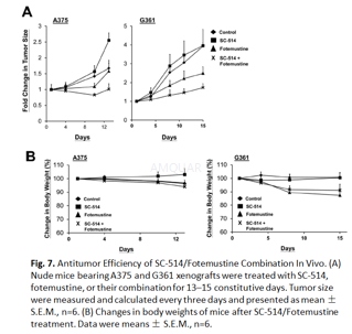
Male nu/nu BALB/c mice (6 weeks old) were maintained in individual ventilated cages in a specific animal handling room. A375 or G361 (5×10⁶) cells were resuspended in 0.1 ml PBS and inoculated subcutaneously into the backs of nude mice and allowed to grow for 7 days. After that, mice were randomly assigned to 4 groups (n=6 for each group) and treated by intraperitoneal injection with 200μl 30% PEG/5% Tween-80 solution as the vehicle control and 25 mg/kg SC-514 and/or 25 mg/kg fotemustine daily for 13–15 consecutive days. Body weight and tumor volume were measured every 3 days. Tumor volumes were determined by a caliper and calculated according to the following formula: (width2×length)/2. At the end of the experiment, mice were sacrificed and tumor xenografts were collected.
-
不同实验动物依据体表面积的等效剂量转换表(数据来源于FDA指南)
|  动物 A (mg/kg) = 动物 B (mg/kg)×动物 B的Km系数/动物 A的Km系数 |
|
例如,已知某工具药用于小鼠的剂量为88 mg/kg , 则用于大鼠的剂量换算方法:将88 mg/kg 乘以小鼠的Km系数(3),再除以大鼠的Km系数(6),得到该药物用于大鼠的等效剂量44 mg/kg。
-
参考文献
[1] Liu Q, Wu H, Chim SM, et al. SC-514, a selective inhibitor of IKKbeta attenuates RANKL-induced osteoclastogenesis and NF-kappaB activation. Biochem Pharmacol. 2013;86(12):1775-1783.
more
分子式
C9H8N2OS2 |
分子量
224.3 |
CAS号
354812-17-2 |
储存方式
﹣20 ℃冷藏长期储存。冰袋运输 |
溶剂(常温)
|
DMSO
53 mg/mL |
Water
<1 mg/mL |
Ethanol
<1 mg/mL |
体内溶解度
-
Clinical Trial Information ( data from http://clinicaltrials.gov )
注:以上所有数据均来自公开文献,并不保证对所有实验均有效,数据仅供参考。
-
相关化合物库
-
使用AMQUAR产品发表文献后请联系我们


