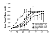
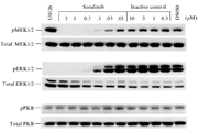
-
生物活性
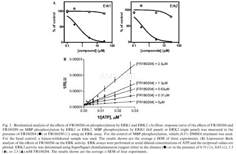
ERK Inhibitor II, FR180204 is a novel ERK-selective inhibitor that can permeate the cell. FR180204 has been reported to inhibit ERK1 (Ki=0.31μM), ERK2 (Ki=0.14 μM), TGFβ-induced AP-1 activation, and acts as a competitive inhibitor of ATP. Displays 30-fold selectivity for ERK over p38α (IC50 = 10 μM); displays no activity against human recombinant MEK1, MKK4, IKKα, PKCα, Src, Syc and PDGFα at concentrations less than 30 μM. Also inhibits TGFβ-induced AP-1 activation in Mv1Lu cells (IC50 = 3.1 μM). Collagen-induced arthritis studies have described that in the presence of FR180204 delayed-type hypersensitivity is weakened and there is a decrease in plasma anti-CII antibody levels. Inhibitor-ERK2 complex studies report that FR180204 shows little activity towards IKKα, MEK1, MKK4, PDGFRα, PKCα, Src, and Syk. ERK Inhibitor II, FR180204 is an inhibitor of p38 α.
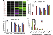
The inhibition of kinases[1]

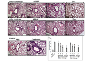
The activity of FR180204 in a cell-based functional assay[1]

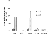
The effects of FR180204 on the viability of human CRC cells[2]

-
体外研究
-
体内研究
悬浮在0.1%甲基纤维素溶液中
-
激酶实验
ERK assay[1]
Nunc-Immuno MaxiSorp plates were coated with 20μg/ml MBP solution in phosphate-buffered saline (PBS). After washing with PBS containing 0.05% Tween 20 (T-PBS), blocking buffer (T-PBS containing 3% BSA) was added to each well and the plates were incubated for 10 min at room temperature. After washing with T-PBS, chemical compounds, ATP and recombinant ERK2 diluted in assay dilution buffer (20 mM Mops, pH 7.2, 25 mMβ-glycerol phosphate, 5 mM EGTA, 1 mM sodium orthovanadate, 1 mM dithiothreitol, and 50μg/ml BSA) and were added to each well. Vehicle groups (containing 0.1% DMSO) and kinase-withdrawal groups were used for the control and basal determinations. After incubation for 1 h at room temperature, plates were washed twice with T-PBS. Anti-phospho MBP antibody (0.2μg/ml) was added to each well, and the plates were incubated for 1 h at room temperature. After washing, anti-mouse HRP-conjugated polyclonal antibodies were added and the plates were incubated for 30 min. SuperSignal chemiluminescent substrate was used for the measurement of HRP activity according to the manufacturers instructions. Prism 4.0 software was used for the Lineweaver–Burk plot analysis, IC50 and Ki determinations.
-
细胞实验
Cell culture[2]
Thehuman CRC DLD‑1 and LoVo cell lineswere cultured in RPMI‑1640 medium containing 10% FBS, 2mM L‑glutamine, 100U/ml penicillin and 100μg/ml streptomycin. The cells were maintained in a humidified atmosphere incubator at 37˚C, with a 5% CO2atmosphere. FR and API‑1 were dissolved in dimethyl sulfoxide (DMSO) to make 1mM stock solutions that were kept at‑20˚C. The stock solutions were freshly diluted with cell culture medium to the required concentration immediately prior to use. The final concentration of DMSO in culture medium during the treatment of cells did not exceed 0.5% (v/v).
Cell viability and apoptotic analyses
To detect the effect of FR and API‑1 on cell viability following treatment, a WST‑1 cell proliferation assay was performed. In brief, DLD‑1 and LoVo cells were seeded into 96‑well plates (1x10⁴ cells/well) containing 100μl of the growth medium in the absence or presence of increasing concentrations of FR (1‑150μM) and API‑1(0.1‑100μM) and then incubated at 37˚C and 5% CO2for 24 and 48 h. At the end of the incubation period, the medium was removed, 100μl WST‑1 was added and the cell solution was incubated at 37˚C for 4 h. Formazan dye produced by viable cells was quantified by measuring absorbance at a wavelength of 450 nm using a microplate reader. All experiments were performed four times and the experiment was repeated twice.

-
动物实验
Animals[3]
Male DBA/1 mice aged 6 weeks were housed in a clean atmosphere with a 12-h light/dark cycle and fed with standard rodent chow ad libitum. They were acclimated for 2 weeks and used at
8 weeks of age.
Induction and evaluation of CIA
Mice were randomized and grouped by body weight immediately before treatment. Bovine CII was dissolved in 0.1 M acetic acid at the concentration of 10 mg/ml and then emulsified in an equal volume of Freund’s complete adjuvant H37Rv. Apart from a naive group, each mouse was immunized with 25μl of the CII emulsion into the tail base, followed by a boost injection 3 weeks after primary injection (day 0). FR180204 and methylprednisolone were ground and suspended in 0.1% methylcellulose solution to a volume of 5 ml/kg. Drugs were given by twice daily intraperitoneal injection from days 0 to 12 in accordance with pharmacokinetic studies with superior area under the curve and Cmax values of i.p. versus s.c. or p.o. administration. The initial administration was 30 min before the boost CII injection. The clinical score of arthritis was obtained by summing the visual severity grade of each limb, scored as follows: 0, no arthritis; 1, one swollen digit; 2, two or more swollen digits or swelling of the entire paw without ankylosis; 3, gross swelling with ankylosis of the paw. Body weight was measured on day 12.
Delayed-type hypersensitivity responses to CII
DBA/1 mice were immunized with 200μg of denatured CII emulsion. CII/phosphate-buffered saline (PBS; 4 mg/ml) was prepared in 0.1-M acetic acid, and then dialyzed against PBS (pH 7.4). The CII/PBS or PBS (25μl) was intradermally injected into the left or right hind paw pad, respectively. The footpad thickness was measuredusing a G-2 dial thickness gauge and represented as the difference in thickness between the injected and the uninjected (control) sides.

-
不同实验动物依据体表面积的等效剂量转换表(数据来源于FDA指南)
|  动物 A (mg/kg) = 动物 B (mg/kg)×动物 B的Km系数/动物 A的Km系数 |
|
例如,已知某工具药用于小鼠的剂量为88 mg/kg , 则用于大鼠的剂量换算方法:将88 mg/kg 乘以小鼠的Km系数(3),再除以大鼠的Km系数(6),得到该药物用于大鼠的等效剂量44 mg/kg。
-
参考文献
[1] Ohori M, Kinoshita T, Okubo M, et al. Identification of a selective ERK inhibitor and structural determination of the inhibitor-ERK2 complex. Biochem Biophys Res Commun. 2005;336(1):357-363.
more
分子式
C18H13N7 |
分子量
327.34 |
CAS号
865362-74-9 |
储存方式
﹣20 ℃冷藏长期储存。冰袋运输 |
溶剂(常温)
|
DMSO
72 mg/mL |
Water
<1 mg/mL |
Ethanol
3 mg/mL |
体内溶解度
-
Clinical Trial Information ( data from http://clinicaltrials.gov )
注:以上所有数据均来自公开文献,并不保证对所有实验均有效,数据仅供参考。
-
相关化合物库
-
使用AMQUAR产品发表文献后请联系我们


