-
生物活性
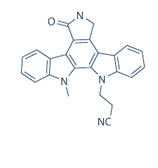
Potent protein kinase C (PKC) inhibitor (IC50 = 7.9 nM). Discriminates between Ca2+-dependent and -independent isoforms of PKC in vitro; selectively inhibits PKCα and PKCβ1 (IC50 values are 2.3 and 6.2 nM respectively).
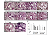
Mean IC50 values of PKC inhibition by GO 6976[1]

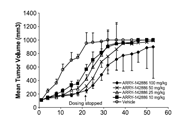
The effect of calcium on PKC inhibition[1]

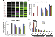
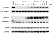
Kinase inhibitory profile of Go6976 in vitro IC50[2]

Cell growth inhibition by Go6976 in human leukemia cell lines harboring FLT3/ITD[2]

Gö6976 blocks cellular CNP-dependent cGMP elevations in 293T-GC-B cells with IC50 of 380 nM.[3]
-
体外研究
-
体内研究
-
激酶实验
In vitro kinase assays[2]
Specific kinase/substrate pairs along with required cofactors were prepared in reaction buffer; 20mM Hepes pH 7.5, 10mM MgCl2, 1mM EGTA, 0.02% Brij35, 0.02mg/ml BSA, 0.1mM Na3VO4, 2mM DTT, 1% DMSO. Compounds were delivered into the reaction, followed ~20 min later by addition of a mixture of ATP and33P ATP to a final concentration of 10mM. Reactions were carried out at 25OC for 120min, followed by spotting of the reactions onto P81 ion exchange filter paper. Unbound phosphate was removed by extensive washing of filters in 0.75% phosphoric acid. After subtraction of background derived from control reactions containing inactive enzyme, kinase activity data were expressed as the percentage of remaining kinase activity in test samples compared to vehicle (dimethyl sulfoxide) reactions. Substrate peptides (single-letter code for amino acids) were: FLT3, Abltide (KKGEAIYAAPFA- NH2); Aurora A and Aurora B, Kemptide (LRRASLG); FGFR3, pEY; JAK2, pEY.
-
细胞实验
Cell culture[4]
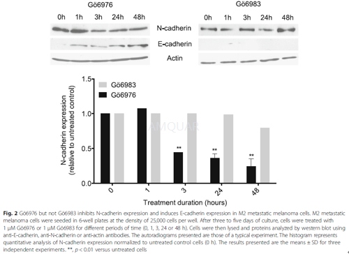
Primary (T1 and I5), their respective lymph-node metastasis (G1 and M2), and the cutaneous metastasis (M4T2) melanoma cell lines were obtained and cultured in RPMI medium supplemented with 10% fetal bovine serum (FBS), 100 units/mL penicillin and 100μg/mL streptomycin (P/S), and 1mM sodium pyruvate (complete medium).
Western blot analysis
Cells were lysed for 20 min at 4 °C in 50 mM Tris–HCl pH 7.4, 150 mM NaCl, 1 mM EDTA, 100 mM sodium fluoride, 10 mM tetra-sodium diphosphate decahydrate, 2 mM sodium orthovanadate, 1 mM phenylmethylsulfonylfluoride, 10 μg/mL aprotinin and 1% Nonidet P-40. Lysates were clarified by centrifugation at 14,000 rpm for 10 min at 4 °C. 30–80 μg of total protein extracts were separated by SDS-PAGE and transferred onto nitrocellulose membranes. These were incubated with the specific antibodies overnight at 4°C and revealed by enhanced chemiluminescence.
-
动物实验
Animals[5]
Balb/c mice (6–8 weeks old; weight range 20–22 g) were obtained and maintained under controlled conditions (22oC, 55% humidity and 12 h day/night rhythm) and fed standard laboratory chow.
Experimental protocol
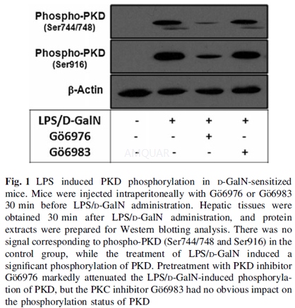
Mice were injected intraperitoneally (i.p.) with Go6976 or Go6983 (2.5 mg/kg, dissolved in DMSO) or equal volume of vehicle 30 min prior to challenge. Mice were challenged i.p. with LPS (50μg/kg) and D-GalN (800 mg/kg). In addition, the doses of Go6976 or Go6983 alone did not induce liver injury, as determined by evaluating liver enzymes, cytokines, and liver histology.
Mice were sacrificed by decapitation at different time points after LPS/D-GalN challenge, and hepatic tissue as well as serum samples were harvested and stored at -80oC for further experiments. Liver samples were harvested 0.5 h after LPS/D-GalN challenge in order to detect levels of phospho-PKD (Ser744/748 and Ser916), phospho-ERK, phospho-JNK and phospho-p38. TNF-a levels in serum and hepatic tissue were measured 1.5 h after LPS/D-GalN challenge. At 6 h after LPS/D-GalN challenge, levels of aminotransferases (AST and ALT) in serum and MPO activity in liver tissue were determined to evaluate hepatic lesion and inflammation; liver tissue was also fixed in formalin for morphological analysis. To determine the effect of PKD inhibition on the outcome of fulminant hepatic failure, lethality (n = 25) was evaluated within 48 h after LPS/D-GalN administration.
Western blotting
The protein of hepatic samples was prepared using the protein extract kit (20 mM Tris, 150 mM NaCl, 1 mM EDTA, 1 mM EGTA, 1% TritonX-100, 2.5 mM sodium pyrophosphate, 1 mM Na3VO4, 1mMβ-glycerolphosphate, 1μg/ml leupeptin and aprotinin). Protein concentrations were determined by a BCA protein assay kit. Protein extracts (40μg) were fractionated on 12% polyacrylamide-sodium dodecyl sulfate (SDS) gel and then transferred to nitrocellulose membrane. The membrane was blocked with 5% (w/v) fat-free milk in Tris-buffered saline (TBS) containing 0.05% Tween 20, followed by incubation with a rabbit primary polyclonal antibody (1:2,000) at 4oC overnight. The membrane was then treated with horseradish peroxidaseconjugated goat anti-rabbit secondary antibody (1:10,000). Antibody binding was visualized with an ECL chemiluminescence system and short exposure of the membrane to X-ray films.
-
不同实验动物依据体表面积的等效剂量转换表(数据来源于FDA指南)
|  动物 A (mg/kg) = 动物 B (mg/kg)×动物 B的Km系数/动物 A的Km系数 |
|
例如,已知某工具药用于小鼠的剂量为88 mg/kg , 则用于大鼠的剂量换算方法:将88 mg/kg 乘以小鼠的Km系数(3),再除以大鼠的Km系数(6),得到该药物用于大鼠的等效剂量44 mg/kg。
-
参考文献
[1] Georg Martiny-Baron MGK, Harald Mischak, Peter M. Blumberg, Georg Kochsll , Hubert Hug, Dieter Marme, Christoph Schachtele. Selective Inhibition of Protein Kinase C Isozymes by the Indolocarbazole GO 6976. J Biol Chem. . 1993;268(13):9194-9197.
[2] Yoshida A, Ookura M, Zokumasu K, Ueda T. Go6976, a FLT3 kinase inhibitor, exerts potent cytotoxic activity against acute leukemia via inhibition of survivin and MCL-1. Biochem Pharmacol. 2014;90(1):16-24.
more
分子式
C24H18N4O |
分子量
378.43 |
CAS号
136194-77-9 |
储存方式
﹣20 ℃冷藏长期储存。冰袋运输 |
溶剂(常温)
|
DMSO
20 mg/mL |
Water
<1 mg/mL |
Ethanol
<1 mg/mL |
体内溶解度
-
Clinical Trial Information ( data from http://clinicaltrials.gov )
注:以上所有数据均来自公开文献,并不保证对所有实验均有效,数据仅供参考。
-
相关化合物库
-
使用AMQUAR产品发表文献后请联系我们


