-
生物活性
MG132 [Z-Leu-Leu-Leu-CHO] is a proteasome inhibitor, used as a tool for perturbing the proteasome-regulated degradation of intracellularproteins.[6][8]
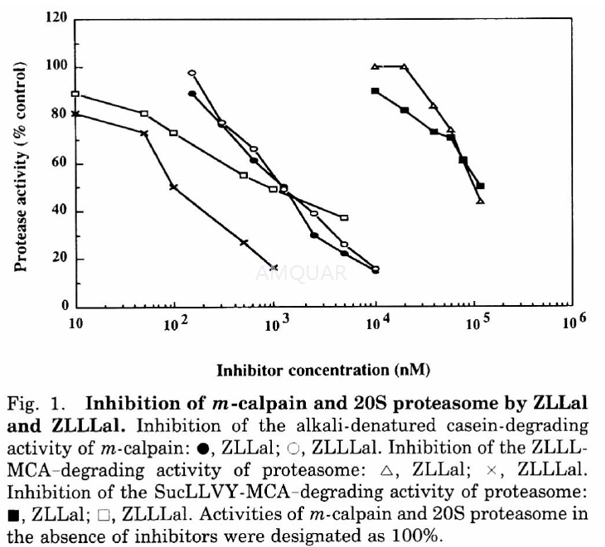
SucLLVY-MCA-degrading activity of proteasome: IC50 = 850 nM; ZLLL-MCA-degrading activity of proteasome: IC50 = 100 nM; calpain: IC50 = 1.2 µM; NF-κB activation: IC50 = 10 µM; IκBα degradation: IC50=10 µM[6][8]
-
体外研究
Proteasome inhibitor MG132 induces selective apoptosis in glioblastoma cells through inhibition of PI3K/Akt and NF-κB pathways, mitochondrial dysfunction, and activation of p38-JNK1/2 signaling[1]. Proteasome inhibitor MG132 significantly blocked pancreatic-cancer-associated angiogenesis through inhibition of NF-κB and NF-κB-dependent proangiogenic gene products VEGF and IL-8[2]. MG-132 sensitizes TRAIL-resistant prostate cancer cells1 by activating the AP-1 family members c-Fos and c-Jun, which, in turn, repress the antiapoptotic molecule c-FLIP(L)[3]. MG132 treatment can improve the developmental potential of Debao porcine SCNT embryos through regulating the expression of genes related to histone acetylation and the processes of ZGA[4]. MG-132 displays 1100 times more effective than ZLLal in inhibiting the ZLLL-MCA-degrading activity of 20S proteasome with IC50 of 100 nM versus 110 μM. MG-132 also inhibits calpain with IC50 of 1.2 μM. MG-132 induces neurite outgrowth in PC12 cells at an optimal concentration of 20 nM, displaying 500 times more potency than ZLLal[5]. MG-132 (10 μM) potently inhibits TNF-α-induced NF-κB activation and IL-8 protein release in A549 cells by inhibition of proteasome-mediated IκBα degradation[6]. MG-132 induces P53 dependent KIM - 2 cell apoptosis through inhibition of 26S proteasome which plays a pivotal role for the degradation pathway in progression through the cell cycle in proliferating cells[7]. Compare with BzLLLCO-CHO, MG-132 shows weaker inhibition of the CT-L and PGPH activities. On contrary, multiple myeloma cells were more sensitive to induction of apoptosis by MG-132 than BzLLLCO-CHO[8].
-
体内研究
1% DMSO+30% polyethylene glycol+1% Tween 80, pH 4
-
激酶实验
Measurement of Inhibitory Activities of ZLLal and ZLLLal (MG132) against m-Calpain and 20S Proteasome.
For the m-calpain inhibitory assay, the 0.5ml reaction mixture contained 0.24% alkali-denatured casein, 28mM 2-mercaptoethanol, 0.94 unit of m-calpain, ZLLal or ZLLLal, 6mM CaCl2, and 0.1M Tris-HC1 (pH 7.5). The reaction was started by the addition of m-calpain solution and stopped by the addition of 0.5ml of 10% trichloroacetic acid after incubation at 30 oC for 15 min. After centrifugation at 1,300 X g for 10 min, the absorbance of the supernatant at 280 nm was measured. The reaction mixture for the 20S proteasome inhibitory assay contained 0.1M Tris-acetate, pH 7.0, 20S proteasome, ZLLal or ZLLLal, and 25 μM substrate dissolved in dimethyl sulfoxide in a final volume of 1ml. After incubation at 37 ℃ for 15 min, the reaction was stopped by the addition of 0.1ml of 10% SDS and 0.9 ml of 0.1M Trisacetate, pH 9.0. The fluorescence of the reaction products was measured. To determine the IC50S against m-calpain and 20S proteasome, various concentrations of the synthetic peptide aldehydes were included in the assay mixture.[5]
-
细胞实验
Cytotoxicity Assay
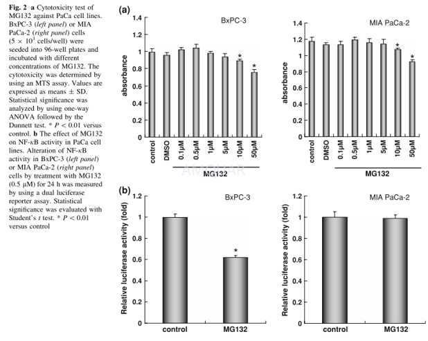
To examine the influence of MG132 on the cytotoxicity against PaCa, we performed Celltiter 96 Aqueous One Solution cell proliferation assay (MTS assay) (Promega, Madison, WI). Cells (5 9 103/well) were seeded in 96-well tissue culture plates and incubated at 37 ℃ with different concentrations of MG132. After 48 h incubation, cell proliferation was measured using anMTSassay kit according to the manufacturer’s instructions. Absorbances were mea- sured using a microplate reader with a test wavelength of 490 nm. Based on the results of these studies, we selected a 0.5 lM concentration of MG132 for further experiments.
Proliferation Assay
HUVECs were seeded at a density of 3 9 103 cells/100 ll into 96-well plates and allowed to adhere overnight. Culture medium was replaced, and the cells were then cultured in medium alone (control) or in medium containing different concentrations of IL-8 or VEGF. After 48 h incubation, cell proliferation was measured by MTS assay according to the manufacturer’s instructions. To examine the effect of PaCa on HUVEC proliferation, we made conditioned medium as follows. BxPC-3 or MIA PaCa-2 cells were seeded at 5 9 106 cells into 100-mm dishes containing medium with 10% FCS and then cultured overnight. Cells were next cultured for 48 h with or without MG132 (0.5 lM). Using this conditioned medium, HUVEC proliferation was determined as described above.
Enzyme-Linked Immunosorbent Assay (ELISA)
BxPC-3 and MIA PaCa-2 cells were seeded at density of 2 9 105 cells/ml into a 24-well plate containing medium with 10% FCS and cultured overnight. The medium was then changed, and cells were cultured for a further 48 h with or without the addition of different concentrations of MG132. The culture medium was then collected and spun in a microcentrifuge at 1,500 rpm for 5 min to remove particles; the supernatants were frozen at -80 ℃ until they were used in the ELISA. The concentrations of VEGF and IL-8 were measured using an ELISA kit (R&D Systems) according to the manufacturer’s instructions.[2]

-
动物实验
Animals and muscle atrophy-induced procedure
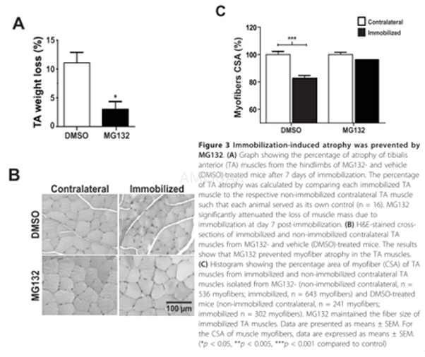
Sixteen-week-old male CD1 mice were used for the experiment. During the immobilization period, the mice were injected subcutaneously with MG132 (7.5 mg/kg/dose) or vehicle (DMSO) twice daily. DMSO containing or not MG132 was diluted in sterile pure corn oil (1:100, injected volume 150 μL). After 7 days, the tibialis anterior (TA) muscles of immobilized and non-immobilized hindlimbs from MG132- and DMSO-treated mice were either harvested or unstapled for remobilization studies.[9]
-
不同实验动物依据体表面积的等效剂量转换表(数据来源于FDA指南)
|  动物 A (mg/kg) = 动物 B (mg/kg)×动物 B的Km系数/动物 A的Km系数 |
|
例如,已知某工具药用于小鼠的剂量为88 mg/kg , 则用于大鼠的剂量换算方法:将88 mg/kg 乘以小鼠的Km系数(3),再除以大鼠的Km系数(6),得到该药物用于大鼠的等效剂量44 mg/kg。
-
参考文献
[1] Zanotto-Filho A, Braganhol E, Battastini AM, Moreira JC. Proteasome inhibitor MG132 induces selective apoptosis in glioblastoma cells through inhibition of PI3K/Akt and NFkappaB pathways, mitochondrial dysfunction, and activation of p38-JNK1/2 signaling. Invest New Drugs. 2012;30(6):2252-2262.
[2] Matsuo Y, Sawai H, Ochi N, et al. Proteasome inhibitor MG132 inhibits angiogenesis in pancreatic cancer by blocking NF-kappaB activity. Dig Dis Sci. 2010;55(4):1167-1176.
[more]
分子式
C26H41N3O5 |
分子量
475.62 |
CAS号
133407-82-6 |
储存方式
﹣20 ℃冷藏长期储存。冰袋运输 |
溶剂(常温)
|
DMSO
100 mM |
Water
partly soluble |
Ethanol
100 mM |
体内溶解度
约5.5 mg/mL
-
Clinical Trial Information ( data from http://clinicaltrials.gov )
注:以上所有数据均来自公开文献,并不保证对所有实验均有效,数据仅供参考。
-
相关化合物库
-
使用AMQUAR产品发表文献后请联系我们


