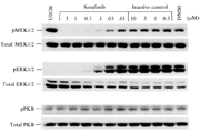
激酶实验
Radioligand Binding Assay[1]
Natalizumab Fab was labeled with 125Iusing IODO-GEN to a specific activity of 134Ci/mmol and concentrated to1.5mg/ml. Jurkat cells (1 x 10⁷/ml) in HBS (20mM HEPES, pH 7.4,137mM NaCl, 5mM KCl, 5.5mM glucose, 10mg/ml of BSA) were treated with 4mM Mn2+and0.4mM Ca2+ (with or without VCAM D1D2) for 30 min at 37 °C. Cellswere aliquoted into 0.6-ml centrifugal tubes (10μl) and put onice. Various concentrations of 125I-natalizumab Fab in 10μlof HBS were added to the cells to give final cation concentrations of 2mM Mn2+ and 0.2mM Ca2+. After 2 h, bound ligand was quantified. Nonspecificbinding was determined in the presence of 100μg/ml of coldnatalizumab IgG for Fab concentrations of 10 nM or lower. At higher Fabconcentrations, the IgG concentration was increased proportionally. Specific bindingwas calculated by subtracting nonspecific Fab binding from total binding.
Competition with increasing concentrationsof non-labeled natalizumab Fab or IgG was performed similarly with 20nM 125I-natalizumabFab. Protein concentrations were determined by measuring A280. Extinctioncoefficients for natalizumab Fab and IgG were calculated from amino acidsequences for the constant regions of human immunoglobulin γ4and κ chains (Uniprot) and the variable regions of natalizumab. Values were76,250 M-1 for Fab fragment and 224,320 M-1 for IgG.

动物实验
Animals and induction of EAE[2]
All experiments were conducted using 6–8 weeksof age adult female SJL mice. Animals were housed and maintained in a controlledenvironment at 22–24oC and 55% humidity, on 12h light/dark cycles andfed with regular rodent’s chow and tap water ad libitum. Animals were handled for1week prior to the beginning of the experiment, in order to reduce the stress inducedby the injections.
For the induction of the relapsing-remittingEAE model, female SJL mice were subcutaneously injected in the hind flanks, with0.2mg of the encephalitogenic proteolipid protein (PLP) 139–151peptide (HSLGKWLGHPDKF),in Complete Freund’s Adjuvant (CFA), on days 0 and 7. Each mouse was also intraperitoneallyinjected with 0.2μg of pertussistoxin on days 0, 1, 7 and 8. Control mice were injectedwith phosphate buffer saline (PBS) in CFA and pertussistoxin. All mice were weighedand examined daily for the clinical signs of EAE, which was scored according tothe following scale: grade 0, no abnormality; grade 1, reduced tail tonus or slightclumsy gait; grade 2, tail atony, moderately clumsy gait, impaired righting ability,or any combination of these signs; grade 3, additional hind limb weakness; grade4,hind limb paralysis and fore limb weakness; grade 5, tetraplegia or mori bund state.Mice developed an acute form of EAE with a peak between days 12 and 14 (grades 1–4),which was termed onset. A phase of complete remission took place between days 19and22, with some mice undergoing are lapse (grades 1 and2) after a variable periodof time (27–31days).
Natalizumabtreatment
In the natalizumab treatment protocol, EAE wasinduced and after appearance of the first clinical signs animals were injected intraperitoneallywith 5mg/Kg of anti-VLA-4antibody (natalizumab) or in the case of control mice withrat IgG. To better represent clinical use, treatment injections were performed duringthe active phase (1, 3 and 5 days after the appearance of the symptoms) (i.e., revertingsymptoms).Animals were sacrificed at day 14 (that corresponds to the onset phasefor animals not treated with natalizumab).
Tissueand CSF collection
Mice were sacrificed during the first peak ofdisease (onset), during the remission phase and in the relapse phase. Mice fromthe control group were sacrificed on day 14, along with mice from the onset group.Animals were anesthetized with ketamine hydrochloride (150mg/Kg) plus medetomidine(0.3mg/Kg), CSF samples were collected from the cisterna magna, and animals weretranscardially perfused with cold saline. CSF pooled samples were checked for bloodcontamination and stored at –80 oC. After perfusion the CP’swere rapidly removed from each mouse ventricle under conventional light microscopy,frozen in dry ice and stored at–80 oC.
For the expression studies, two sets of animalsfrom each experimental group (control, onset, remission and relapse) were prepared:one for the microarray analysis containing 3 separate pooled CP samples for onsetand remission phase and 2-pooled CP for the relapse phase (from three animals each)and another to the quantitative Real Time-Polymerase Chain Reaction (qRT-PCR) containingat least from 5 pools of CP (from three animals each).
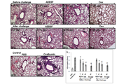
Immunohistochemistryand immunofluorescence analyses
Animals were transcardially perfused withsaline, under anesthesia, at the various time points mentioned before. Afterperfusion, brains were removed from the skull, immediately embedded in Tissue-Tekoptimal cutting temperature compound and kept frozenat –20oC. Brainswere sectioned in serial 20μm cryostat coronal sections, which were then fixed with4% PFA in PBS. After antigen retrieval with citrate buffer 10mM, the sections wereprobed with the primary antibody, anti-mouse lipocalin-2/neutrophil gelatinase-associatedlipocalin (LCN2/NGAL) (1:400), diluted in PBS0 3% Triton X-100 (PBS-T), and 0.4%bovine serum albumin. Afterwards the sections were incubated with biotinylated anti-goatsecondary antibody and then with streptavidin peroxidase conjugate (ABC kit). Reactionwas developed with3, 3’-diaminobenzidine tetrahydrochloride hydrate (DAB) and sectionsstained with hematoxylin. Alternatively, for immunofluorescent staining, sectionswere incubated with goat anti-mouse LCN2/NGAL in conjunction with: rabbit anti-glialfibrillary acidic protein (anti-GFAP) (1:200) to label astrocytes, rat anti-mouseLy-6Gto label neutrophils (1:200), mouse anti-CNPase to label oligodendrocytes (1:100),mouse anti-NeuN (1:100) to label neurons and rabbit anti-Iba1 (1:200) to label microglia.The appropriate secondary fluorescent antibodies diluted 1:500 in PBS-T were used:anti-goat Alexa 488, anti-rabbit and anti-mouse Alexa 594, anti-rat Alexa 547. Foreach analysis, sections from 3–5 different animals pergroup were used. The cellnucleus was stained using 4’, 6-diamidino-2-phenylindole (DAPI). Samples were analyzedusing optical or confocal microscopes.
参考文献
[1] Yu, Y.; Schurpf, T.; Springer, T. A., How natalizumab binds and antagonizes alpha4 integrins. J Biol Chem 2013, 288 (45), 32314-25.
[2] Marques, F.; Mesquita, S. D.; Sousa, J. C.; Coppola, G.; Gao, F.; Geschwind, D. H.; Columba-Cabezas, S.; Aloisi, F.; Degn, M.; Cerqueira, J. J.; Sousa, N.; Correia-Neves, M.; Palha, J. A., Lipocalin 2 is present in the EAE brain and is modulated by natalizumab. Frontiers in Cellular Neuroscience 2012, 6.
分子式
|
分子量
|
CAS号
|
储存方式
-80 ℃长期储存。干冰运输 |
溶剂(常温)
|
DMSO
|
Water
|
Ethanol
|
体内溶解度